. 2003 Mar 30;9(1):7-19.
doi: 10.1177/159101990300900102. Epub 2004 Oct 22.

Effects of endothelial injury on the rate of thrombus organization in canine carotid arteries occluded with microcoils

Affiliations

Effects of endothelial injury on the rate of thrombus organization in canine carotid arteries occluded with microcoils

T Abruzzo et al. Interv Neuroradiol. .

Abstract

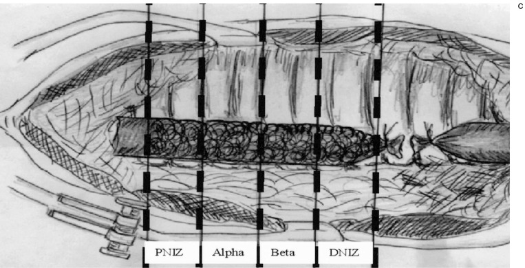
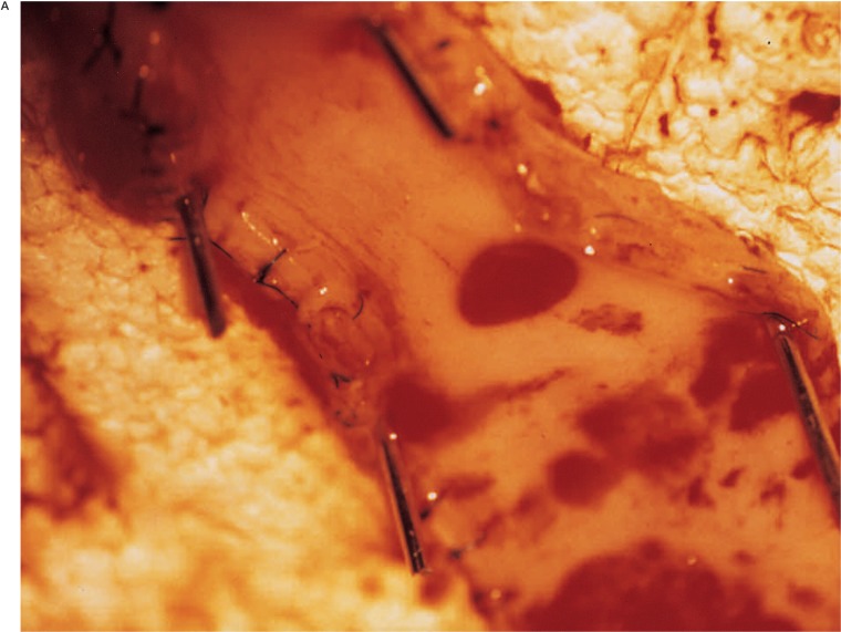
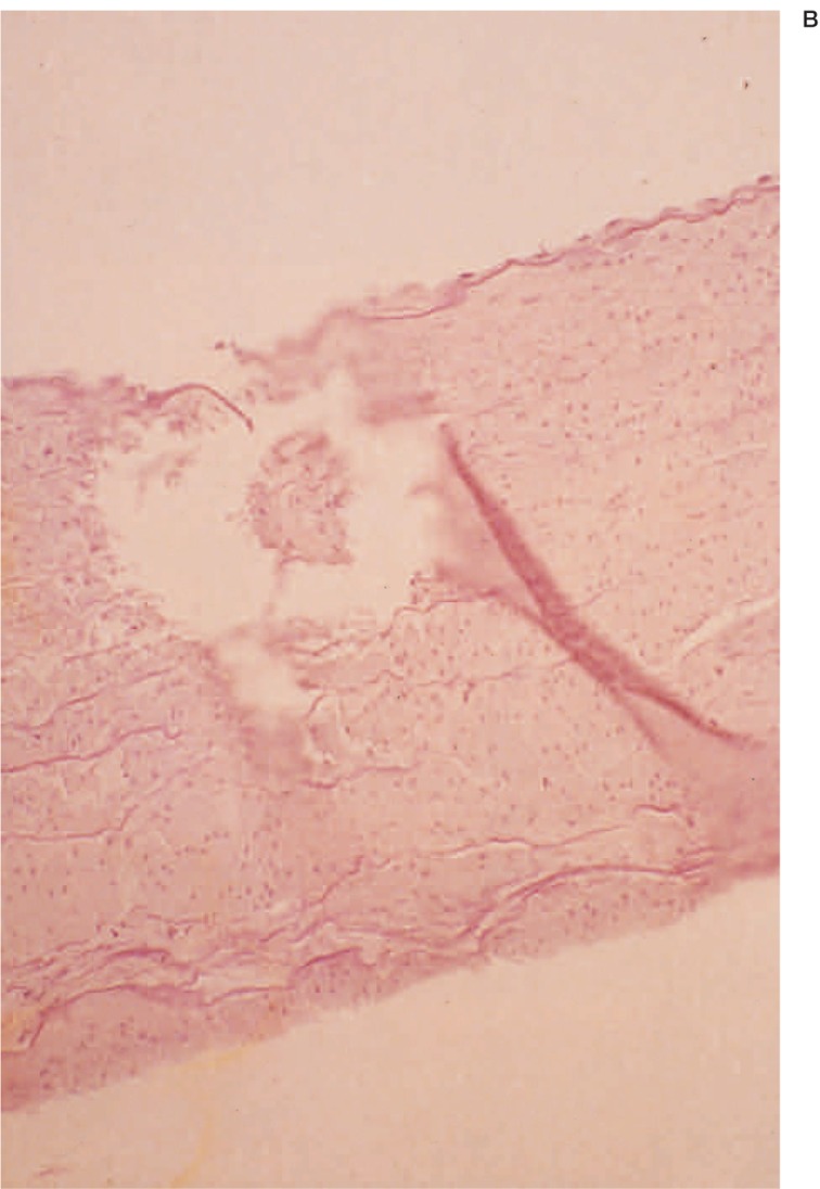
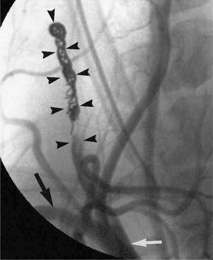
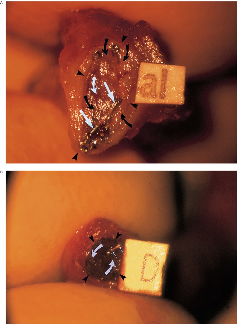
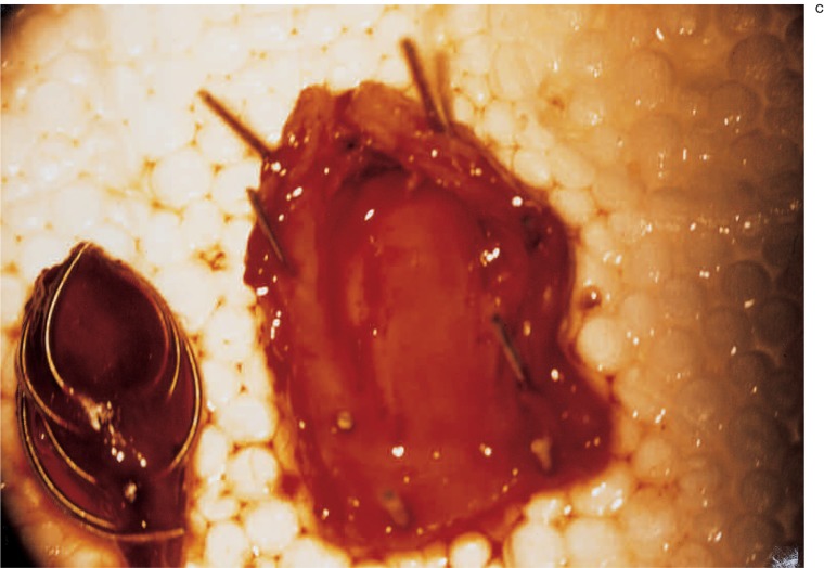
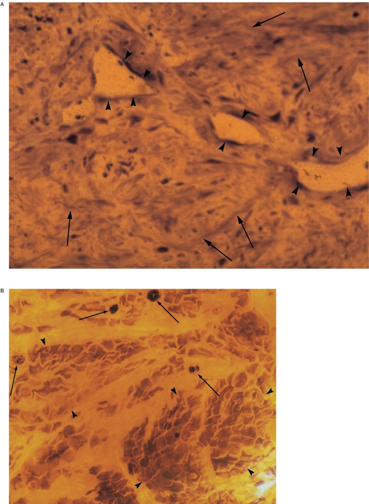
Thrombus organization in canine carotid arteries occluded with platinum microcoils was studied to determine if endothelial injury created with a Xenon Chloride Excimer Laser (XEL) could acclerate endovascular fibrosis. Ten common carotid artery stumps were created in ten dogs. Each of four stumps were schematically divided into four longitudinally contiguous injury zones (thermal ablation injury, non-ablative injury, proximal and distal non-injury zones) to test the effects of ablative and non-ablative injury and to establish a set of internal controls that would account for proximity to circulating blood at the ostium of the occluded artery. Following XEL irradiation of the endothelium through an arteriotomy, each stump was embolized with microcoils. Four control stumps were subjected to sham laser procedures, and embolized in an identical fashion. Two additional stumps were embolized in the absence of sham surgery. Angiographic, gross and histologic analysis was performed after four weeks. Specimens of freshly clotted whole blood mixed with microcoils were used as an additional control. In irradiated stumps and non-irradiated stumps (sham and embolization only), angiography revealed no evidence of coil compaction or recanalization. In all irradiated stumps the thermal ablation zone contained fibrous tissue and neovascularity without unorganized thrombus. The other zones in the irradiated stumps were indistingnishable from each other and from all zones in the non-irradiated sham stumps, containing primarily unorganized thrombus. Stumps embolized in the absence of sham surgery were filled with material that was grossly and microscopically identical to specimens of freshly clotted whole blood containing microcoils. The results indicate that thermal ablation injury of the endothelium accelerates thrombus organization in canine carotid arteries occluded with platinum microcoils.


Figures

Figure 1
Figure 1
B) Technique of laser irradiation and coil embolization. Following closure of the arteriotomy, a 6 french guide catheter (curved black arrow) is introduced into the open end of the CCA stump, and the distal ostium of the stump is cinched down around the tip of the catheter with a temporary ligature. A tracker 18 microcatheter (long straight black arrows) is directed proximally through the guide catheter and the stump is progressively occluded from proximal to distal with microcoils (short straight black arrows) by incrementally withdrawing the microcatheter. C) Technique of laser irradiation and coil embolization.Following removal of both catheters, the distal end of the stump is suture ligated, and the proximal clip is removed to reestablish flow.Proximal Non-Injury Zone (PNIZ), Distal Non-Injury Zone (DNIZ).
Figure 1
Figure 1
B) Technique of laser irradiation and coil embolization. Following closure of the arteriotomy, a 6 french guide catheter (curved black arrow) is introduced into the open end of the CCA stump, and the distal ostium of the stump is cinched down around the tip of the catheter with a temporary ligature. A tracker 18 microcatheter (long straight black arrows) is directed proximally through the guide catheter and the stump is progressively occluded from proximal to distal with microcoils (short straight black arrows) by incrementally withdrawing the microcatheter. C) Technique of laser irradiation and coil embolization.Following removal of both catheters, the distal end of the stump is suture ligated, and the proximal clip is removed to reestablish flow.Proximal Non-Injury Zone (PNIZ), Distal Non-Injury Zone (DNIZ).
Figure 2
Figure 2
A) Pathology of thermal ablation injury. Endoluminal surface of a canine common carotid artery viewed en-face (original magnification × 16). The arterial lumen is opened along a longitudinal incision and the edges are tacked back by dissection pins. A subacute thermal ablation injury harvested 48 hours after creation, as part of the preliminary dosimetry study, demonstrates a large halo of subintimal hemorrhage extending beyond the boundaries of the irradiated endothelial surface. B) Pathology of thermal ablation injury. Longitudinal section of an acute thermal ablation injury harvested immediately after creation, as part of the preliminary dosimetry study, shows sharply circumscribed crater edges and microdissections extending into the arterial wall (hematoxylin and eosin;original magnification × 10).
Figure 2
Figure 2
A) Pathology of thermal ablation injury. Endoluminal surface of a canine common carotid artery viewed en-face (original magnification × 16). The arterial lumen is opened along a longitudinal incision and the edges are tacked back by dissection pins. A subacute thermal ablation injury harvested 48 hours after creation, as part of the preliminary dosimetry study, demonstrates a large halo of subintimal hemorrhage extending beyond the boundaries of the irradiated endothelial surface. B) Pathology of thermal ablation injury. Longitudinal section of an acute thermal ablation injury harvested immediately after creation, as part of the preliminary dosimetry study, shows sharply circumscribed crater edges and microdissections extending into the arterial wall (hematoxylin and eosin;original magnification × 10).
Figure 3
Figure 3
Arteriography. Right carotid arteriogram obtained immediately after coil embolization demonstrates ≥ 90% occlusion of a sham CCA stump, with uniform coil packing throughout. Black arrowheads indicate the right common carotid artery stump. White arrow indicates the rachiocephalic artery, and black arrow indicates right subclavian artery.
Figure 4
Figure 4
Gross pathology. A) Trans-axial cross-section through the alpha zone of a common carotid artery stump four weeks after laser irradiation and coil embolization (original magnification × 10). Endoluminal coils (straight white arrows) are encased in fibrous scar tissue (curved black arrows) and granulation tissue (curved white arrows). The lumen (border indicated by black arrowheads) is deformed into an eccentric configuration due to scar tissue contraction. B) Trans-axial cross-section through the distal non-injury zone of a common carotid artery stump 4 weeks after laser irradiation and coil embolization (original magnification × 10). Endoluminal coils (straight white arrows) are suspended in unorganized red thrombus (curved white arrows). Note that the rounded configuration of the lumen (borders indicated by black arrowheads) is preserved. C) Group 3 right common carotid artery stump 4 weeks after coil embolization (original magnification × 16). The arterial lumen has been opened along its longitudinal axis, and a mass of red thrombus intermixed with coils has been removed.
Figure 4
Figure 4
Gross pathology. A) Trans-axial cross-section through the alpha zone of a common carotid artery stump four weeks after laser irradiation and coil embolization (original magnification × 10). Endoluminal coils (straight white arrows) are encased in fibrous scar tissue (curved black arrows) and granulation tissue (curved white arrows). The lumen (border indicated by black arrowheads) is deformed into an eccentric configuration due to scar tissue contraction. B) Trans-axial cross-section through the distal non-injury zone of a common carotid artery stump 4 weeks after laser irradiation and coil embolization (original magnification × 10). Endoluminal coils (straight white arrows) are suspended in unorganized red thrombus (curved white arrows). Note that the rounded configuration of the lumen (borders indicated by black arrowheads) is preserved. C) Group 3 right common carotid artery stump 4 weeks after coil embolization (original magnification × 16). The arterial lumen has been opened along its longitudinal axis, and a mass of red thrombus intermixed with coils has been removed.
Figure 5
Figure 5
Histopathology. A) Trans-axial cross-section through the alpha zone of a common carotid artery stump four weeks after laser irradiation and coil embolization (toluidine blue; original magnification × 40). The lumen is filled with connective tissue (collagen fibers indicated by straight black arrows) that is penetrated by neovascularity (black arrowheads). B) Longitudinal cross-section through the central region of a sham lumen shows poorly organized clot stained with hemosiderin. The arrows indicate multinucleate giant cells (toludine blue; original magnification × 40). Black arrowheads indicate erythrocytes.

Similar articles

References

    1. Byrne J, Sohn M, Molyneux A. Five year experience in using coll embolization for ruptured intracranial aneurysms: outcomes and incidence of late rebleeding. Journal of Neurosurgery. 1999;90:656–663. - PubMed
    1. Cognard C, Weill A, et al. Long-term angiographic follow-up of 169 intracranial berry aneurysms occluded with detachable coils. Radiology: 1999;212:348–356. - PubMed
    1. Molyneux A, Ellison D, et al. Histological findings in giant aneurysms treated with Guglielmi detachable coils: report of two cases with autopsy correlation. Journal of Neurosurgery: 1996;83:129–132. - PubMed
    1. Manabe H, Fujita S, et al. Re-rupture of coil-em-bolized aneurysms during long-term observation. Journal of Neurosurgery. 1998;88:1096–1098. - PubMed
    1. Castro E, Fortea F, et al. Long term histopathologic findings in two cerebral aneurysms embolized with Guglielmi detachable coils. American Journal of Neuroradiology. 1999;20:549–552. - PMC - PubMed

LinkOut - more resources