

doi: 10.1177/159101990300900406.
Epub 2004 Oct 22.
Secondary spontaneous thrombosis of a giant aneurysm located distally on a feeding artery after embolization of an associated arteriovenous malformation
Affiliations
- PMID: 20591316
- PMCID: PMC3547378
- DOI: 10.1177/159101990300900406
Item in Clipboard
Secondary spontaneous thrombosis of a giant aneurysm located distally on a feeding artery after embolization of an associated arteriovenous malformation
Interv Neuroradiol.
.
Abstract
Giant aneurysm located in the distal part of the feeding artery associated with a cerebral arteriovenous malformation is rare and the treatment is clinically challenging. We report the spontaneous and complete thrombosis of a flow-related giant aneurysm immediate up-stream to a cerebral arteriovenous malformation by embolization of that malformation alone in a patient presenting with complex partial seizure and no history of intracranial haemorrhage. We obviated the need to directly intervene on the giant aneurysm, thus reducing unnecessary procedure related risks to the patient. Follow up one year later confirms the thrombosis and show shrinkage of the mass. The patient is asymptomatic.
Figures

A) Frontal and B) lateral views of digital subtraction cerebral angiograms demonstrating the architecture of the cerebral AVM and the associated up-stream giant aneurysm. C) Medial and D) lateral 3 dimensional reconstruction images of the cerebral AVM and the associated giant aneurysm.

Lateral view of the venous phase of the cerebral angiogram after embolization of the cerebral AVM, showing blood-contrast level within the giant aneurysm, indicating immediate cessation of blood flow within the aneurismal lumen.

CT image of the brain obtained 48 hours post-embolization of the cerebral AVM, indicating complete thrombosis of the giant aneurysm. Note the hyperdense glue-lipiodol mixture within the AVM nidus.

A) Frontal and B) lateral views, obtained two weeks post-embolization of the cerebral AVM. The giant aneurysm was no longer opacified with contrast. A small residual AVM was still present.
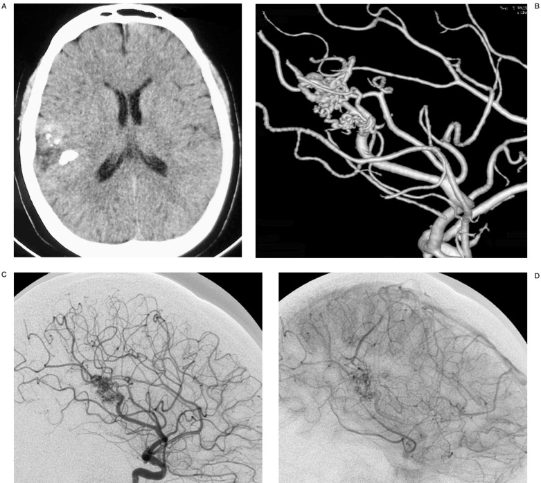
A) CT control 1 year later. B-D) lateral views. The giant aneurysm is no longer opacified and has completely shrunked. A small residual AVM is still present.
References
-
- Anderson RM, Blacwood W. The association of arteriovenous angioma and saccular aneurysm of the arteries of the brain. J Pathol Bacteriol. 1959;77:101–110. - PubMed
-
- Batjer H, Suss RA, Samson D. Intracranial arteriovenous malformations associated with aneurysms. Neurosurgery. 1986;18:29–35. - PubMed
-
- Arai H, Sugiyama Y, et al. Multiple intracranial aneurysms and vascular malformations in an infant. Case report. J Neurosurg. 1972;37:357–360. - PubMed
-
- Aarabi B, Chambers J. Giant thrombosed aneurysm associated with an arteriovenous malformation. Case report. J Neurosurg. 1978;49:278–282. - PubMed
LinkOut - more resources
Full Text Sources
