. 2003 Dec 20;9(4):407-24.
doi: 10.1177/159101990300900413. Epub 2004 Oct 22.

Dural Sinus Malformations (DSM) with Giant Lakes, in Neonates and Infants. Review of 30 Consecutive Cases

Affiliations

Dural Sinus Malformations (DSM) with Giant Lakes, in Neonates and Infants. Review of 30 Consecutive Cases

M Barbosa et al. Interv Neuroradiol. .

Abstract

Abstract: Background and Purpose. Dural Arteriovenous Shunt (DAVS) in children include Dural sinus malformation (DSM), infantile and adult types. They are rare and seldom reported. Our purpose was to highlight the angiographic features of the DSM sub group for prognosis of clinical evolution and outcome and to lay guidelines for management.

Methods: From a dedicated neurovascular data bank, there were 52 cases of arteriovenous dural shunts in children from 1985 to 2003. Of these, there were 30 patients with DSM, which we analysed the various angioarchitecture, presentation and neurological outcome. Children clinical status was evaluated and scored at admission and follow up. Results. There was an overall male dominance of 2:1. Antenatal diagnosis was obtained in 8/30 (26.7%) cases. Mean age of diagnosis was 5 months. Mean age at first consultation was 8.7 months. No patient was diagnosed during childhood. The most common clinical presentations were macrocrania 76.7%, seizures 23.3% and mental retardation 23.3%. In 14/30 (35.7%) of the patients, the therapeutic decision was to manage conservatively; in 5/14 (30.7%) with predictable favourable evolution and in 9/14 (64.3%) with irreversible poor neurological outcome. In the remaining 16/30 (53.3%) patients, endovascular treatment was performed. In 12/16 (75.0%) patients the neurological outcome was good, 3/16 (18.8%) patients had unfavourable evolution despite embolization. There was no morbidity mortality related to the procedures themselves. 1/16 (6.3%) patient was lost to follow-up. Overall 12/29 (45.8%) patients had an unfavourable neurological outcome with 11 patients dead and 1 with severe neurological deficit. In the surviving group of children, 17/18 (94.4%) have a good neurological outcome; in 10/18 (55.5%) the lesion is morphologically excluded. Conclusion. DSM is rare disease with high mortality. They usually proceed to either total or partial spontaneous thrombosis before the age of 2 thus compromising normal cerebral venous drainage. DSM away from the torcular, good cavernous sinus, cavernous capture of sylvian veins, absence of pial veins, straight sinus or superior sagital sinus (SSS) reflux and absence of jugular bulb dysmaturation represent factors of good prognosis. Such patients will highly benefit for endovascular treatment. In partial endovascular approach the aim being is to separate the brain drainage from DSM drainage. This will be achieved by the transarterial approach to the associated mural arterio-venous shunts (AVS) and by disconnecting the pial reflux by transvenous route.


Figures

Scheme 1
Scheme 1
Figure 1
Figure 1
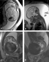
Antenatal MRI diagnosis of DSMs with (A,B) and without (C, D) evidence of intraluminal thrombosis.
Figure 2
Figure 2
(Case 23) A-C) Antenatal MRI diagnosis of DSM. D-F) post natal evidence of intra luminal thrombosis. G-I) complete remodelling.
Figure 3
Figure 3
A-B) Neonatal MRI diagnosis of DSM away from the torcular. C-D) post natal spontaneous thrombosis.
Figure 4
Figure 4
(Case 19) A-C) neonatal MRI showing torcular DSM. D-F) 7 months FU MRI shows multiple cavernomas with haemorrhagic changes in the right periventricular region and enlargement of the DSM to the Right transverse sinus.
Scheme 2
Scheme 2
Figure 5
Figure 5
(Case 14) A-D) Mural AVS from the R. intra cavernous ICA. No deep vein seen, bilateral cavernous sinus capture, posterior fossa congestion. Jugular veins patent bilaterally downstream jugular foramina. E-G) Mural AVS from the ascending pharyngeal and middle meningeal arteries;-Torcular involvement-Straight sinus and SSS reflux-Left Jugular bulb dysmaturation. H) retrograde injection of the straight sinus prior to its trans venous disconnection. Note the opacification of the cavernous drainage of the deep system. I-M) Following trans venous occlusion of the straight sinus, Left transverse and SSS with coils (I), few AVS were embolised leading to substantial reduction in flow into the malformed sinus (J-M). N-Q) 4 years FU angiogram shows remodeling of the cerebral venous drainage. Note the bilateral cerebellar DVA draining into the Left petrous vein and subsequently into the cavernous sinus and downstream the bulb occlusion into the Left jugular vein.
Figure 5
Figure 5
(Case 14) A-D) Mural AVS from the R. intra cavernous ICA. No deep vein seen, bilateral cavernous sinus capture, posterior fossa congestion. Jugular veins patent bilaterally downstream jugular foramina. E-G) Mural AVS from the ascending pharyngeal and middle meningeal arteries;-Torcular involvement-Straight sinus and SSS reflux-Left Jugular bulb dysmaturation. H) retrograde injection of the straight sinus prior to its trans venous disconnection. Note the opacification of the cavernous drainage of the deep system. I-M) Following trans venous occlusion of the straight sinus, Left transverse and SSS with coils (I), few AVS were embolised leading to substantial reduction in flow into the malformed sinus (J-M). N-Q) 4 years FU angiogram shows remodeling of the cerebral venous drainage. Note the bilateral cerebellar DVA draining into the Left petrous vein and subsequently into the cavernous sinus and downstream the bulb occlusion into the Left jugular vein.
Figure 6
Figure 6
(Case 15) Remodelling following trans arterial embolization of mural AVS.

References

    1. Lasjaunias P. Interventional Neuroradiology Management. Berlin Heidelberg: Springer - Verlag; 1997. Vascular diseases in neonates, infants and children; pp. 321–371.
    1. Morita A, Meyer FB, et al. Childhood dural arteriovenous fistulae of the posterior dural sinuses: three case reports and literature review. Neurosurgery. 1995;37:1193–1200. - PubMed
    1. Lasjaunias P, Magufis G, et al. Anatomoclinical Aspects of Dural Arteriovenous Shunts in Children: Review of 29 cases. Interventional Neuroradiology. 1996;2:179–191. - PubMed
    1. Albright AL, Latchaw RE, Price RA. Posterior dural arteriovenous malformation in infancy. Neurosurgery. 1983;13:129–135. - PubMed
    1. Garcia-Monaco R, Rodesch G, et al. Multifocal dural arteriovenous shunts in children. Childs Nerv Syst. 1991;7:425–431. - PubMed

LinkOut - more resources