. 2010 Oct;68(10):2412-21.
doi: 10.1016/j.joms.2009.09.058. Epub 2010 Jun 29.

Comparison of actual surgical outcomes and 3-dimensional surgical simulations

Affiliations

Comparison of actual surgical outcomes and 3-dimensional surgical simulations

Scott Tucker et al. J Oral Maxillofac Surg. 2010 Oct.

Abstract

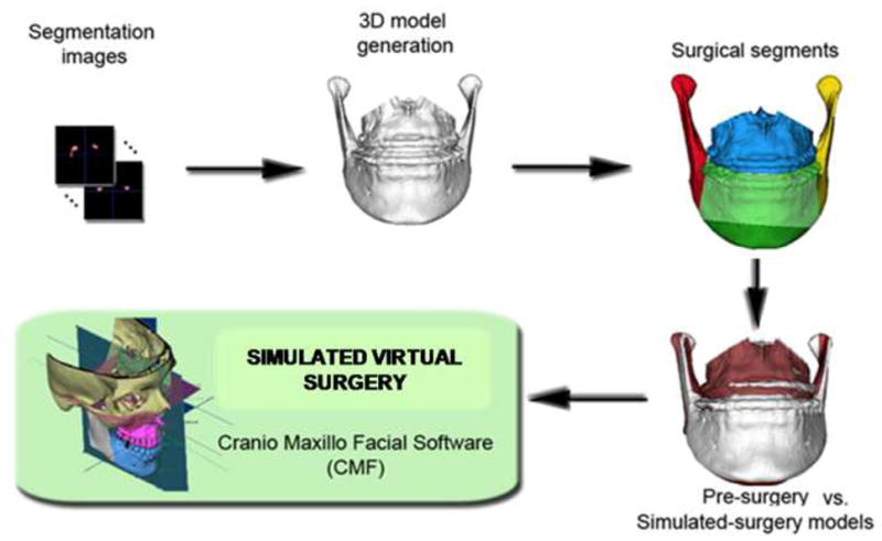
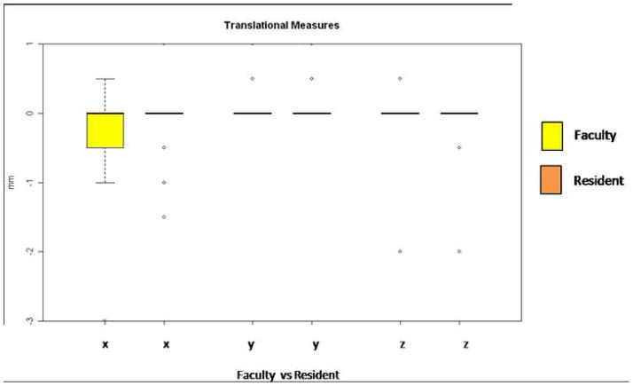
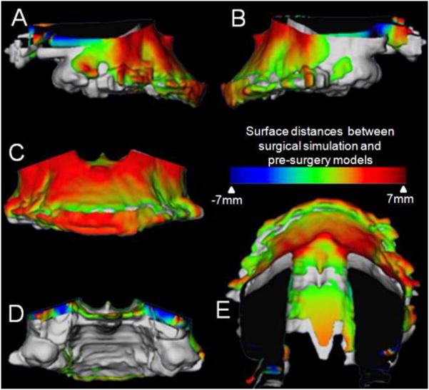
Purpose: The advent of imaging software programs has proved to be useful for diagnosis, treatment planning, and outcome measurement, but precision of 3-dimensional (3D) surgical simulation still needs to be tested. This study was conducted to determine whether the virtual surgery performed on 3D models constructed from cone-beam computed tomography (CBCT) can correctly simulate the actual surgical outcome and to validate the ability of this emerging technology to recreate the orthognathic surgery hard tissue movements in 3 translational and 3 rotational planes of space.

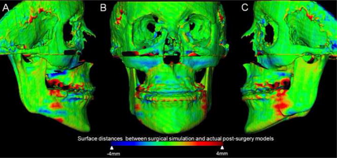
Materials and methods: Construction of pre- and postsurgery 3D models from CBCTs of 14 patients who had combined maxillary advancement and mandibular setback surgery and 6 patients who had 1-piece maxillary advancement surgery was performed. The postsurgery and virtually simulated surgery 3D models were registered at the cranial base to quantify differences between simulated and actual surgery models. Hotelling t tests were used to assess the differences between simulated and actual surgical outcomes.

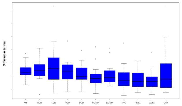
Results: For all anatomic regions of interest, there was no statistically significant difference between the simulated and the actual surgical models. The right lateral ramus was the only region that showed a statistically significant, but small difference when comparing 2- and 1-jaw surgeries.

Conclusions: Virtual surgical methods were reliably reproduced. Oral surgery residents could benefit from virtual surgical training. Computer simulation has the potential to increase predictability in the operating room.


Figures

Figure 1
Figure 1
Sequence of image analysis procedures used for virtual surgical simulation: After segmentation of anatomic structures, i.e. outlining the shape of structures visible in the cross-sections of a CBCT volumetric dataset, the virtual cuts were performed. For each patient, simulated surgery outcomes were created, to compare to presurgery and actual surgery models. Virtual cuts matched clinical osteotomy segments that in this example were: chin, left ramus, right ramus, mandibular body and/or maxillary body. The virtual surgical segments were then displaced to determine if virtual surgery performed on the Cone beam CT surface models can correctly simulate the actual surgical outcome.
Figure 2
Figure 2
Superimposition of virtual surgery models and post surgery models of a patient treated with maxillary advancement and mandibular setback. A, Right lateral view, B, Frontal view, and C, Left lateral view. Color maps demonstrate the location, direction, and magnitude of the differences between these models. Note that in the maxilla and mandible except for areas of surgical cuts the surface distances between simulated and actual surgery models are close to 0mm (green).
Figure 3
Figure 3
The differences between virtual and actual post-surgery models are shown below. The x axis shows the 11 regions of interest and the y axis shows the difference in mm between the two images. All regions of interest except the left lateral maxilla showed a mean and median difference less than the 0.5 mm spatial resolution of the acquired image. (Ant = Anterior Maxilla, RLat = Right lateral maxilla, LLat = Left lateral maxilla, RCon = Right condyle, LCon = Left condyle, RLRam = Right lateral ramus, LLRam = Left lateral ramus, AntC = Anterior Corpus of the mandible, RLatC = Right lateral corpus of the mandible, LLatC = Left lateral corpus of the mandible, Chin = Chin)
Figure 4
Figure 4
Translational movements of the right and left lateral rami during mandibular setback surgery. The faculty member operated on the right side and is always shown in the left of the paired columns (yellow bloxplot; x, y, z coordinates). The resident operated on the left side and is always shown on the right of the paired columns (orange bloxplots; x, y, z coordinates). Directions of movement in mm: x coordinates, (+)left/(−)right movement; y coordinates (+)anterior/(−)posterior; and z coordinates (+)superior/(−)inferior movement.
Figure 5
Figure 5
Rotational movements of the right and left lateral rami during mandibular setback surgery. The faculty member operated on the right side and is always shown in the yellow of the paired columns. The resident operated on the left side and is always shown on the orange of the paired columns. Amount of rotation in degrees is shown: (+) signifies a clockwise rotation and (−) signifies a counterclockwise rotation. Column X: Axial plane or Pitch, Column Y: Sagittal plane or Yaw and Column Z: Coronal plane or Roll.
Figure 6
Figure 6
Example of a maxillary impaction case in which surgical simulation helped plan areas and amount of bone removal for impaction. Superimposition of maxillary segment of virtual surgery models and pre-surgery models of patients treated with maxillary advancement and impaction. A, Right lateral view, B, Left lateral view, C, Frontal view, D, Posterior view and E, Superior view. The grey image is the pre-surgery model and the image with the color map is the post virtual (simulated) surgery image. Color maps demonstrate the location, direction, and magnitude of the differences between these models. Note the dark blue area in the posterior part of the maxilla indicating that 7mm of posterior bone removal will be necessary during the surgery.

References

    1. Loh S, Yow M. Computer prediction of hard tissue profiles in orthognathic surgery. Int J Adult Orthodon Orthognath Surg. 2002;17(4):342–7. - PubMed
    1. Loh S, Heng JK, Ward-Booth P, Winchester L, McDonald F. A radiographic analysis of computer prediction in conjunction with orthognathic surgery. Int J Oral Maxillofac Surg. 2001;30(4):259–63. - PubMed
    1. Mankad B, Cisneros GJ, Freeman K, Eisig SB. Prediction accuracy of soft tissue profile in orthognathic surgery. Int J Adult Orthodon Orthognath Surg. 1999;14(1):19–26. - PubMed
    1. Pektas ZO, Kircelli BH, Cilasun U, Uckan S. The accuracy of computer-assisted surgical planning in soft tissue prediction following orthognathic surgery. Int J Med Robot. 2007;3:64–71. - PubMed
    1. Smith JD, Thomas PM, Proffit WR. A comparison of current prediction imaging programs. Am J Orthod Dentofacial Orthop. 2004;125(5):527–36. - PMC - PubMed

Publication types