

Review
doi: 10.1016/j.jjcc.2010.05.008.
Epub 2010 Jun 29.
Patent foramen ovale and stroke
Affiliations
- PMID: 20591626
- PMCID: PMC3723385
- DOI: 10.1016/j.jjcc.2010.05.008
Item in Clipboard
Review
Patent foramen ovale and stroke
J Cardiol.
2010 Sep.
Abstract
The presence of a patent foramen ovale has been found to be associated with an increased risk of ischemic stroke of otherwise unknown origin (cryptogenic stroke). The present article will review the evidence regarding this association, the technical aspects of PFO detection, and the preventive options to decrease the risk of recurrent cerebral events.
Copyright © 2010 Japanese College of Cardiology. Published by Elsevier Ltd. All rights reserved.
Figures
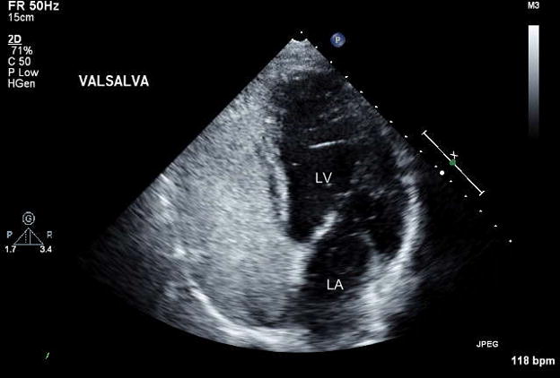
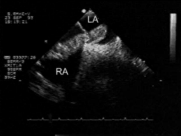
Example of PFO detection by transthoracic echocardiography (TTE) with contrast injection. Microbubbles are visualized filling the right-sided chambers and into the left atrium (LA) and left ventricle (LV).
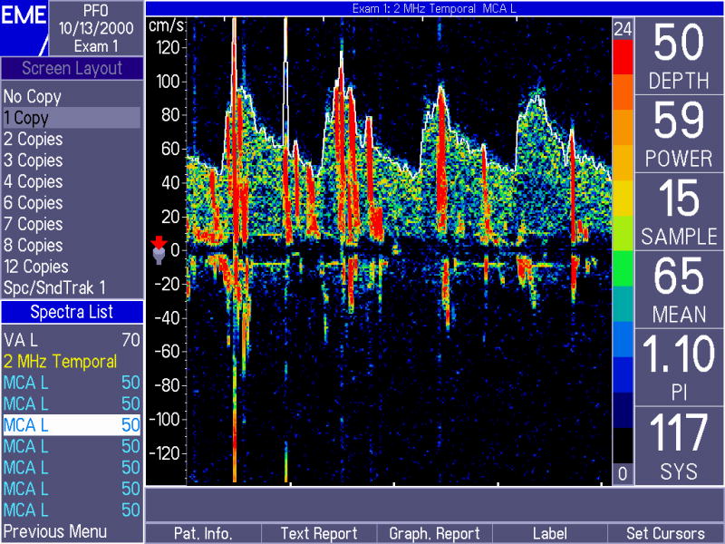
Detection of microbubbles by transcranial Doppler (TCD) in the middle cerebral artery of a patient with a PFO. Microbubbles are visualized as spikes superimposed to the normal blood flow
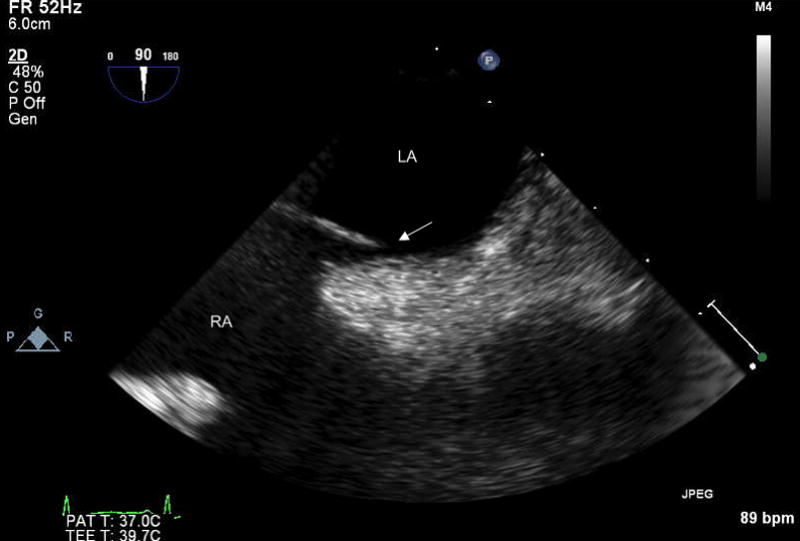
Direct visualization of PFO by TEE. A separation (arrow) is seen between septum primum and septum secundum. LA = left atrium; RA = right atrium
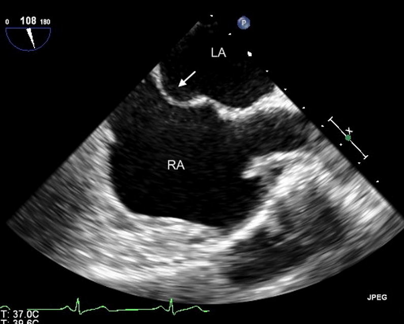
Visualization of atrial septal aneurysm (ASA) by TEE. Protrusion of the atrial septum towards the right atrium is visible (arrow). LA = left atrium; RA = right atrium
Visualization by TEE of large thrombus crossing the PFO (arrows). LA = left atrium; RA = right atrium
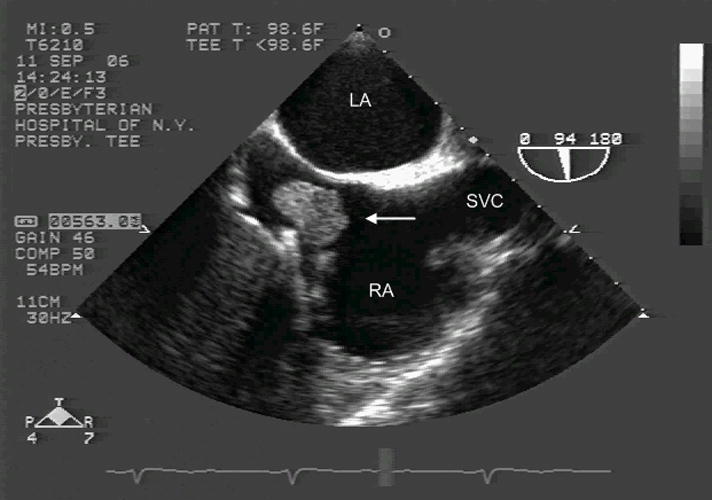
Visualization by TEE of large thrombus (arrow) on the Eustachian valve. LA = left atrium; RA = right atrium; SVC = superior vena cava
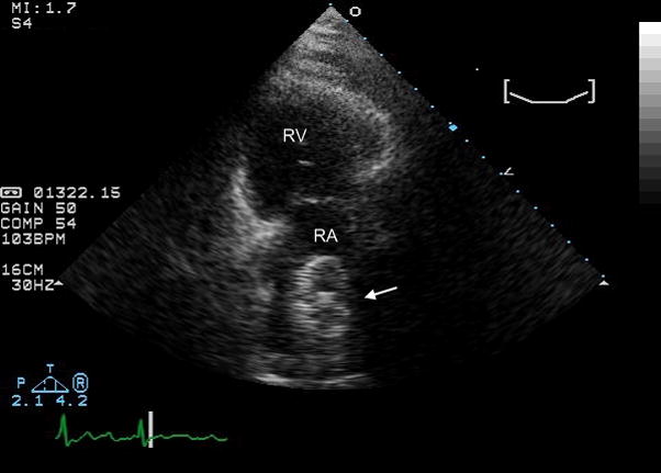
Visualization by TTE (long axis view) of a PFO closing device. RV = right ventricle; RA = right atrium
References
-
- Penther P. Patent foramen ovale: an anatomical study. Apropos of 500 consecutive autopsies. Arch Mal Coeur Vaiss. 1994 Jan;87(1):15–21. - PubMed
-
- Schroeckenstein RF, Wasenda GJ, Edwards JE. Valvular competent patent foramen ovale in adults. Minn Med. 1972 Jan;55(1):11–3. - PubMed
-
- Hagen PT, Scholz DG, Edwards WD. Incidence and size of patent foramen ovale during the first 10 decades of life: an autopsy study of 965 normal hearts. Mayo Clin Proc. 1984 Jan;59(1):17–20. - PubMed
-
- Meissner I, Khandheria BK, Heit JA, Petty GW, Sheps SG, Schwartz GL, Whisnant JP, Wiebers DO, Covalt JL, Petterson TM, Christianson TJ, Agmon Y. Patent foramen ovale: innocent or guilty? Evidence from a prospective population-based study. J Am Coll Cardiol. 2006 Jan 17;47(2):440–5. - PubMed
-
- Rodriguez CJ, Homma S, Sacco RL, Di Tullio MR, Sciacca RR, Mohr JP. Race-ethnic differences in patent foramen ovale, atrial septal aneurysm, and right atrial anatomy among ischemic stroke patients. Stroke. 2003 Sep;34(9):2097–102. - PubMed
Publication types
MeSH terms
Grants and funding
LinkOut - more resources
Full Text Sources
Medical
