Review
. 2010 Aug;13(4):459-65.
doi: 10.1016/j.mib.2010.05.015. Epub 2010 Jun 28.

Parasites in motion: flagellum-driven cell motility in African trypanosomes

Affiliations
Review

Parasites in motion: flagellum-driven cell motility in African trypanosomes

Kent L Hill. Curr Opin Microbiol. 2010 Aug.

Abstract

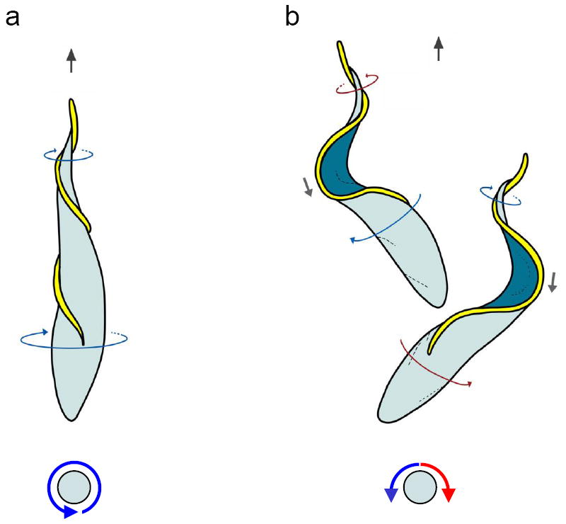
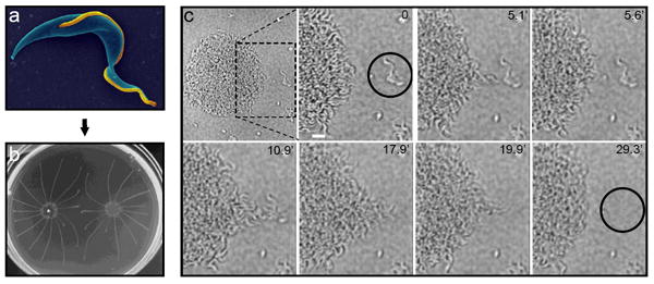
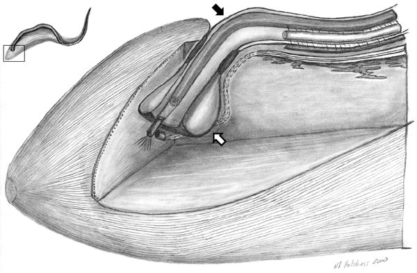
Motility of the sleeping sickness parasite, Trypanosoma brucei, impacts disease transmission and pathogenesis. Trypanosome motility is driven by a flagellum that harbors a canonical 9+2 axoneme, together with trypanosome-specific elaborations. Trypanosome flagellum biology and motility have been the object of intense research over the last two years. These studies have led to the discovery of a novel form of motility, termed social motility, and provided revision of long-standing models for cell propulsion. Recent work has also uncovered novel structural features and motor proteins associated with the flagellar apparatus and has identified candidate signaling molecules that are predicted to regulate flagellar motility. Together with earlier inventories of flagellar proteins from proteomic and genomic studies, the stage is now set to move forward with functional studies to elucidate molecular mechanisms and investigate parasite motility in the context of host-parasite interactions.


Figures

Figure 1
Figure 1. Tryps Rock
Cartoons of T. brucei cells show the classical (a) and bihelical (b) model for T. brucei motility. Direction of cell propulsion is with the flagellum tip leading (black arrows, top). Top panels show view from above the cell and bottom panels show the end-on view, looking at the cell posterior. In panel (a), classical model, a left-handed helical flagellum (yellow) drives rotation of the cell body counterclockwise, looking from the cell posterior toward anterior (blue arrows). In panel (b), bihelical model, the flagellum (yellow) alternately generates right-handed (red arrows) and left-handed (blue arrows) helical waves that propagate from tip to base and drive cell motion with the flagellum tip leading. Between helical segments of opposite handedness the flagellum forms a kink (gray arrows) that propagates in the direction opposite cell movement. The direction of helix rotation changes simultaneously with the change in helix handedness, causing the cell to rock back and forth along its long axis (red and blue arrows), while continuing to move forward. A helix is a chiral form, meaning that a mirror-image of the form cannot be superimposed on the original. Your hand is an example of a chiral object. The handedness of a helix can be determined by wrapping one’s hand around the helix with the thumb aligned along the long axis, then tracing the helix with your finger tips in the direction the thumb is pointing (Supplemental Figure 1). For a given helix, this is possible only with the right or left hand, not both, thus defining the “handedness”. In the examples shown, thin blue arrows indicate flagellum sections with left-handed chirality and thin red arrows indicate flagellum sections having a right-handed chirality. Adapted from [7], with permission.
Figure 2
Figure 2. Social motility reveals that trypanosomes sense, communicate and cooperate
Panel (a) shows an electron micrograph of a procyclic T. brucei cell from suspension culture, pseudo-colored with the flagellum in gold. Panel (b) shows social motility colonies six days post-inoculation on semisolid agarose plates, with characteristic radial projections that migrate outward and avoid other parasites. Panel (c) shows a time-lapse series of the early stages of social motility. Parasites at the colony perimeter, termed “scouts” migrate out and back from the colony. When scouts identify external parasites (black circle in second panel), they return to the colony and direct coordinate movement of cells in the colony outward at this position to recruit the external parasites into the colony. Scale bar is 20 um. Timestamps are indicated in each panel. See also Supplemental Movie 1. Panels a and c are adapted from [5] and [35], respectively, with permission.
Figure 3
Figure 3. The trypanosome flagellar pocket
Cut-away, three-dimensional view of the posterior region of the T. brucei cell, illustrating the flagellar pocket (white arrow) and flagellum (black arrow). The flagellar pocket forms from an invagination of the cell surface membrane at the position where the flagellum (black arrow) emerges from the cytoplasm. The flagellum and flagellar pocket are intimately connected and flagellum motility is hypothesized to influence traffic into and out of the flagellar pocket [41]. The inset, upper left, shows a schematic of the T. brucei cell, with a boxed region to indicate the position of the cutaway view. Adapted from [45], with permission.

Comment in

Similar articles

Cited by

References

    1. Van Den Abbeele J, et al. Trypanosoma brucei spp. development in the tsetse fly: characterization of the post-mesocyclic stages in the foregut and proboscis. Parasitology. 1999;118(Pt 5):469–78. - PubMed
    1. Vickerman K, et al. Biology of African trypanosomes in the tsetse fly. Biol Cell. 1988;64(2):109–19. - PubMed
    1. Ginger ML, Portman N, McKean PG. Swimming with protists: perception, motility and flagellum assembly. Nat Rev Microbiol. 2008;6(11):838–50. - PubMed
    1. Ralston KS, Hill KL. The flagellum of Trypanosoma brucei: new tricks from an old dog. Int J Parasitol. 2008;38(8-9):869–84. - PMC - PubMed
    1. Ralston KS, et al. The Trypanosoma brucei flagellum: moving parasites in new directions. Annual Review of Microbiology. 2009;63:335–62. - PMC - PubMed

Publication types

Substances