. 2010 Nov;38(20):7167-78.
doi: 10.1093/nar/gkq588. Epub 2010 Jun 30.

Class-specific restrictions define primase interactions with DNA template and replicative helicase

Affiliations

Class-specific restrictions define primase interactions with DNA template and replicative helicase

Marilynn A Larson et al. Nucleic Acids Res. 2010 Nov.

Abstract

Bacterial primase is stimulated by replicative helicase to produce RNA primers that are essential for DNA replication. To identify mechanisms regulating primase activity, we characterized primase initiation specificity and interactions with the replicative helicase for gram-positive Firmicutes (Staphylococcus, Bacillus and Geobacillus) and gram-negative Proteobacteria (Escherichia, Yersinia and Pseudomonas). Contributions of the primase zinc-binding domain, RNA polymerase domain and helicase-binding domain on de novo primer synthesis were determined using mutated, truncated, chimeric and wild-type primases. Key residues in the β4 strand of the primase zinc-binding domain defined class-associated trinucleotide recognition and substitution of these amino acids transferred specificity across classes. A change in template recognition provided functional evidence for interaction in trans between the zinc-binding domain and RNA polymerase domain of two separate primases. Helicase binding to the primase C-terminal helicase-binding domain modulated RNA primer length in a species-specific manner and productive interactions paralleled genetic relatedness. Results demonstrated that primase template specificity is conserved within a bacterial class, whereas the primase-helicase interaction has co-evolved within each species.


Figures
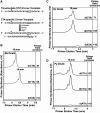
Figure 1.
Figure 1.
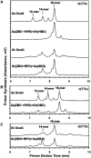
Trinucleotide initiation specificity for primases from B. anthracis, Y. pestis and P. aeruginosa. (A) Schematic of primase activity assay using the 23-mer trinucleotide-specific template where XYZ is the trinucleotide of interest (upper sequence) and with RNA primer synthesis initiating from the middle nucleotide of d(CTA) (lower sequence). The initiation trinucleotide in the ssDNA sequence is underlined, the full-length template-derived primer product is italicized and the unique guanine used to determine the initial nucleotide copied in the RNA primer is denoted with an asterisk. (B–D) Initiation specificity of DnaG primase on various trinucleotide-specific templates and detection of RNA primer products by denaturing HPLC. Chromatograms show the synthesis of RNA primers by B. anthracis (Ba) primase, Y. pestis (Yp) primase and P. aeruginosa (Pa) primase on 23-mer templates containing the recognition trinucleotides d(CTA), d(TTA) or d(CTG), as well as a representative negative template d(ATA). The elution of full-length 16-mer primers initiating from the middle nucleotide in the initiation trinucleotides d(CTA), d(CTG) or d(TTA) are shown and were based on the respective RNA standard. Each increment on the y-axis is equivalent to 5 mV absorbance at 260 nm.
Figure 2.
Figure 2.
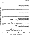
Contribution of the primase HBD on initiation specificity. (A–C) Comparison of primer synthesis by wild-type DnaG from S. aureus (Sa) and E. coli (Ec) to chimeric primases in which the HBD was exchanged. Primases were tested with the 23-mer template containing (A) d(CTA), (B) d(TTA) or (C) d(CTG) as designated in the upper right corner. RNA primers were visualized by denaturing HPLC and their nucleotide lengths were determined by comparison to 12-mer, 14-mer and 16-mer RNA size markers as indicated. Y-axis increments are each equal to 5 mV.
Figure 3.
Figure 3.
Key residues in the primase ZBD determine initiation specificity. (A) Denaturing HPLC analyses of RNA primers synthesized by the ZBD-swapped chimeric primase compared to wild-type primase from S. aureus (Sa) or E. coli (Ec) on templates containing d(CTA), d(TTA) or d(CTG). The 23-mer ssDNA template containing the specific trinucleotide is indicated on the right, with bold bars denoting common templates. Primer lengths are based on RNA size markers as shown. (B) Sequence alignment of the primase N-terminal ZBD from Gamma-proteobacteria and Bacilli Firmicutes with known trinucleotide initiation specificity. The ZBD residues are numbered above the alignment and are based on S. aureus DnaG (bottom sequence). Primases within the same bacterial class are bracketed on the left. Secondary structural elements are indicated above, and the unexposed inner (i) or exposed outer (o) amino acids are denoted below the relevant residues, as previously proposed (4). Open arrowhead indicates the location of the conserved cysteines and histidine. Solid arrow denotes the mutated S. aureus ZBD residues and asterisks indicate the mutated amino acids that confer trinucleotide specificity shown in (C). Identical residues conserved in all six (blue), five (green) or four (cyan) ZBDs are indicated. Conserved Gamma-proteobacteria (yellow) or Bacilli Firmicutes (red) ZBD residues are denoted. Abbreviations are Yp, Y. pestis; Ec, E. coli; Pa, P. aeruginosa; Gs, G. stearothermophilus; Ba, B. anthracis; and Sa, S. aureus. (C) Representative chromatograms showing RNA primer synthesis by the modified S. aureus primase with a single (R32K, L37C, I56F and C57Y) or a double (I56F/C57Y) ZBD mutation, as well as by wild-type DnaG from S. aureus (Sa) and E. coli (Ec). Priming assays were performed with the 23-mer template containing either d(CTA) or d(CTG) designated with an A or G, respectively, on the right. Primer lengths are indicated and each y-axis increment is equivalent to 10 mV.
Figure 4.
Figure 4.
Full-length chimeric primases interact in trans on class-associated trinucleotides. (A and B) Representative chromatograms comparing RNA primer production on the Gamma-proteobacterial initiation trinucleotide d(CTG) by the chimeric primase comprising the S. aureus ZBD and E. coli RPD+HBD (A) or the chimera with the E. coli ZBD and S. aureus RPD+HBD (B). Priming reactions contained 3.6 µM chimeric primase. (C and D) Results of protein-mixing assays with the ZBD-swapped chimeras shown in (A) and (B) at a final concentration of (C) 1.8 µM or (D) 3.6 µM with the d(CTG)-containing 23-mer template. Y-axis increments are each equivalent to 5 mV.
Figure 5.
Figure 5.
Cross-stimulation of primase activity by replicative helicases. (A–G) Unrooted phylogenetic trees based on sequence identities for DnaG primase and replicative helicase and their associated domains from the Bacilli Firmicutes S. aureus (Sa), B. anthracis (Ba) and G. stearothermophilus (Gs) and the Gamma-proteobacteria E. coli (Ec), Y. pestis (Yp) and P. aeruginosa (Pa). Phylogenetic trees for (A) full-length primase, (B) full-length helicase, (C–E) individual primase domains, the (F) helicase N-terminal domain (NTD) and (G) C-terminal domain (CTD) are shown. (H) RNA primer synthesis by DnaG primase from the Firmicutes B. anthracis and G. stearothermophilus in the absence or presence of a Firmicute replicative helicase at stoichiometric concentrations. Reactions were incubated with the 23-mer template containing d(CTA) and either 0.3 µM B. anthracis (Ba) DnaG (top four chromatograms) or 0.9 µM G. stearothermophilus (Gs) DnaG (bottom four chromatograms) without or with 0.1 µM or 0.3 µM of the designated Firmicute helicase hexamer, respectively. Bacillus anthracis and G. stearothermophilus DnaG activity assays contained either no helicase (–) or B. anthracis (Ba), S. aureus (Sa) or G. stearothermophilus (Gs) helicase as indicated on the right. (I) RNA primer synthesis by DnaG primase from the Proteobacteria E. coli and Y. pestis in the absence or presence of a Proteobacteria replicative helicase at stoichiometric concentrations. Reactions were incubated with the 23-mer template containing d(CTG) and either 1.8 µM E. coli (Ec) DnaG (top four chromatograms) or 0.6 µM Y. pestis (Yp) DnaG (bottom four chromatograms) without or with 0.6 µM or 0.2 µM of the designated Proteobacteria helicase hexamer, respectively. E. coli and Y. pestis DnaG activity assays contained either no helicase (–) or E. coli (Ec), Y. pestis (Yp), or P. aeruginosa (Pa) helicase as denoted on the right. Each y-axis increment is equivalent to 10 mV.
Figure 6.
Figure 6.
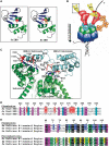
Regulation of primer synthesis by critical DnaG primase and replicative helicase residues. (A) Comparison of the S. aureus (Sa) and E. coli (Ec) primase ZBD models based on consensus structures obtained for A. aeolicus and G. stearothermophilus (PDB codes 2AU3 and 1D0Q, respectively). Two key residues in the β4 strand responsible for trinucleotide specificity are highlighted in red (Ile56) and cyan (Cys57) for the Firmicute S. aureus (left ZBD) and orange (Phe58) and purple (Tyr59) for the Proteobacteria E. coli (right ZBD). The zinc ion is shown as a gray sphere. All molecular structures were generated with the PyMOL molecular graphics software (http://pymol.sourceforge.net). (B) Model of trans interaction between primase ZBD (yellow) with adjacent RPD active site (red) on the class-associated trinucleotide d(CTA). Ribbon structure of boxed ZBD is shown in 6A. Arrow indicates direction of replication fork movement. The primase HBD (cyan) is shown interacting with replicative helicase N-terminal domain (green) that is covalently linked to the C-terminal ATPase domain (blue). SSB proteins (orange) are not shown to scale. (C) Interface between C1 and C2 subdomains of the primase HBD (cyan) and N-terminal domain (NTD) of two replicative helicase protomers (green) from B. anthracis. Models are based on G. stearothermophilus structures (PDB code 2R6C) and numbering refers to the B. anthracis proteins. Residues unique (underlined) and conserved in B. anthracis and G. stearothermophilus that potentially contribute to the productive cross-interaction between DnaG (red) and helicase (blue) are shown. (D) Multiple sequence alignment of the HBD region shown in (C) from the Bacilli Firmicutes B. anthracis (Ba), G. stearothermophilus (Gs) and S. aureus (Sa). Residues unique to B. anthracis and G. stearothermophilus are highlighted in red. Asterisk identifies residues underlined in (C). Identical (cyan) and similar (purple) amino acids conserved in all three Firmicutes are shown. (E) Multiple sequence alignments of the N-terminal region of helicase shown in (C) from the Bacilli Firmicutes B. anthracis (Ba), G. stearothermophilus (Gs) and S. aureus (Sa) and the Gamma-proteobacteria E. coli (Ec), Y. pestis (Yp) and P. aeruginosa (Pa). Identical amino acids unique to B. anthracis and G. stearothermophilus (blue), as well as E. coli and Y. pestis (yellow) are shown. Asterisk indicates residues underlined in (C). Amino acids conserved in either Firmicutes or Proteobacteria (green) and identical (black) or similar (purple) residues conserved in both phyla are shown.

Similar articles

Cited by

References

    1. Kornberg A, Baker T. DNA Replication. 2nd edn. Freeman: San Francisco; 1992.
    1. Okazaki R, Okazaki T, Sakabe K, Sugimoto K, Sugino A. Mechanism of DNA chain growth: I. Possible discontinuity and unusual secondary structure of newly synthesized chains. Proc. Natl Acad. Sci. USA. 1968;59:598–605. - PMC - PubMed
    1. Wu CA, Zechner EL, Reems JA, McHenry CS, Marians KJ. Coordinated leading- and lagging-strand synthesis at the Escherichia coli DNA replication fork: V. Primase action regulates the cycle of Okazaki fragment synthesis. J. Biol. Chem. 1992;267:4074–4083. - PubMed
    1. Pan H, Wigley DB. Structure of the zinc-binding domain of Bacillus stearothermophilus DNA primase. Structure. 2000;8:231–239. - PubMed
    1. Griep MA, Lokey ER. The role of zinc and the reactivity of cysteines in Escherichia coli primase. Biochemistry. 1996;35:8260–8267. - PubMed

Publication types