. 2010 Aug;61(13):3647-62.
doi: 10.1093/jxb/erq178. Epub 2010 Jun 30.

Revealing diversity in structural and biochemical forms of C4 photosynthesis and a C3-C4 intermediate in genus Portulaca L. (Portulacaceae)

Affiliations

Revealing diversity in structural and biochemical forms of C4 photosynthesis and a C3-C4 intermediate in genus Portulaca L. (Portulacaceae)

Elena V Voznesenskaya et al. J Exp Bot. 2010 Aug.

Abstract

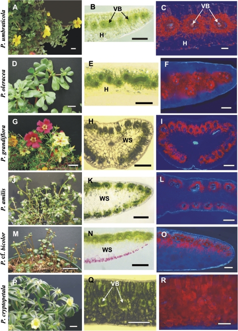
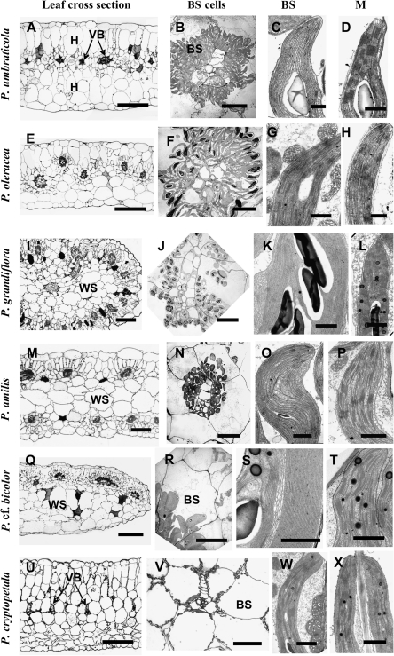
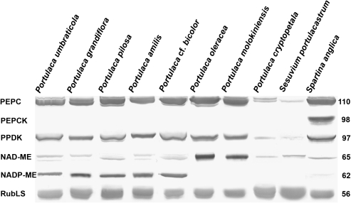
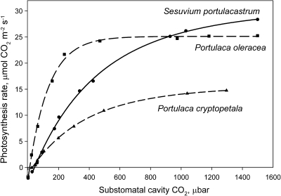
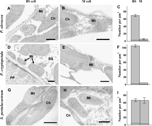
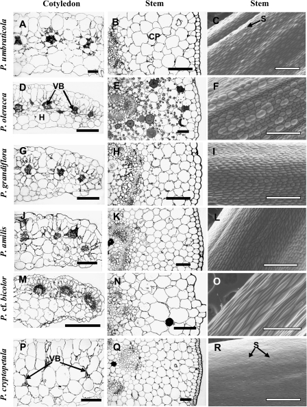
Portulacaceae is one of 19 families of terrestrial plants in which species having C(4) photosynthesis have been found. Representative species from major clades of the genus Portulaca were studied to characterize the forms of photosynthesis structurally and biochemically. The species P. amilis, P. grandiflora, P. molokiniensis, P. oleracea, P. pilosa, and P. umbraticola belong to the subgenus Portulaca and are C(4) plants based on leaf carbon isotope values, Kranz anatomy, and expression of key C(4) enzymes. Portulaca umbraticola, clade Umbraticola, is NADP-malic enzyme (NADP-ME)-type C(4) species, while P. oleracea and P. molokiniensis in clade Oleracea are NAD-ME-type C(4) species, all having different forms of Atriplicoid-type leaf anatomy. In clade Pilosa, P. amilis, P. grandiflora, and P. pilosa are NADP-ME-type C(4) species. They have Pilosoid-type anatomy in which Kranz tissues enclose peripheral vascular bundles with water storage in the centre of the leaf. Portulaca cf. bicolor, which belongs to subgenus Portulacella, is an NADP-ME C(4) species with Portulacelloid-type anatomy; it has well-developed Kranz chlorenchyma surrounding lateral veins distributed in one plane under the adaxial epidermis with water storage cells underneath. Portulaca cryptopetala (clade Oleracea), an endemic species from central South America, was identified as a C(3)-C(4) based on its intermediate CO(2) compensation point and selective localization of glycine decarboxylase of the photorespiratory pathway in mitochondria of bundle sheath cells. The C(4) Portulaca species which were examined also have cotyledons with Kranz-type anatomy, while the stems of all species have C(3)-type photosynthetic cells. The results indicate that multiple structural and biochemical forms of C(4) photosynthesis evolved in genus Portulaca.


Figures

Fig. 1.
Fig. 1.
General view of Portulaca species (left panels), hand-made leaf cross-sections (middle panels), and distribution of chlorenchyma under the fluorescent microscope (right panels). (A–C) Portulaca umbraticola; (B) distribution of vascular bundles (VBs) in the median paradermal plane; (C) red fluorescence from chlorenchyma is distributed around each VB. (D–F) Portulaca oleracea; (E) zig-zag pattern in the distribution of VBs with their positioning in two paradermal levels, (F) fluorescent chlorenchyma surrounds the VBs. (G–I) Portulaca grandiflora and (J–L) P. amilis; (H, K) VBs are distributed around the leaf periphery, (I, L) chlorenchymatous cells are mostly on the outer side of the VBs. (M–O) Portulaca cf. bicolor; (N) VBs are only on the adaxial side, (O) chlorophyll fluorescence occurs on the adaxial and abaxial sides of VBs. (P–R) Portulaca cryptopetala; (Q) C3-like dorsoventral type of anatomy, (R) chlorenchyma is distributed evenly in leaf section. H, hypoderm; VB, vascular bundles; WS, water storage tissue. Scale bars: 2 cm for A, D, G, J, M, P; 500 μm for B, E, H, K, N, Q; 250 μm for C, F, I, L, O, R.
Fig. 2.
Fig. 2.
Light microscopy of leaf cross-sections, electron microscopy of bundle sheath (BS) cells and of chloroplasts and mitochondria in chlorenchyma cells in Portulaca species. (A–D) Portulaca umbraticola. (E–H) Portulaca oleracea. (I–L) Portulaca grandiflora. (M–P) Portulaca amilis. (Q–T) Portulaca cf. bicolor. (U–X) Portulaca cryptopetala. A, E, I, M, Q, U left panels: light microscopy. B, F, J, N, R, V: BS cells with centripetal positioning of organelles surrounding VBs. BS chloroplasts: (C, K, O, S) grana-deficient (G, W) with well-developed grana and numerous mitochondria. M chloroplasts: (D, L, P, T, X) with well-developed grana and (H) deficient in grana. BS, bundle sheath; H, hypoderm; M, mesophyll; VB, vascular bundle; WS, water storage tissue. Scale bars: 250 μm for A, E, I, M, Q, U; 20 μm for B, F, J, N, R; 100 μm for V; 1 μm for C, D, G, H, K, L, O, P, S, T, W, X.
Fig. 3.
Fig. 3.
Western blots for C4 enzymes and Rubisco from total soluble proteins extracted from leaves of eight Portulaca species, the C3 species Sesuvium portulacastrum, and the C4 PEP-CK species Spartina anglica. Blots were probed with antibodies raised against PEPC, PPDK, NAD-ME, NADP-ME, PEP-CK, and Rubisco, respectively. Numbers on the left indicate molecular mass in kilodaltons. Subtle differences in blot intensity might occur due to differences in antigenicity between species.
Fig. 4.
Fig. 4.
Rates of CO2 fixation in response to varying intercellular levels of CO2 at 21% O2, 25 °C, and a PPFD of 900 μmol quanta m−2 s−1 in Portulaca cryptopetala, P. oleracea, and Sesuvium portulacastrum. The results represent the average from measurements of the response to changes in CO2 (from ambient to low, and low to high) from separate measurements on 2–3 leaves from different plants.
Fig. 5.
Fig. 5.
Electron microscopy of in situ immunolocalization of glycine decarboxylase (GDC) in chlorenchyma cells of P. oleracea (A, B), P. cryptopetala (D, E), and Sesuvium portulacastrum (G, H), and graphs showing the density of labelling for GDC in BS versus M mitochondria (right panels C, F, and I). (A, D, G) Immunolabelling of GDC in bundle sheath cell mitochondria. (B, E) Lack of immunolabelling for GDC in M cells of two Portulaca species, and (H) showing the presence of gold particles in M mitochondria in S. portulacastrum. (C, F, I) In all graphs the x-axis represents the number of gold particles per μm2 of mitochondrial area; for each cell type 10–60 cell fragments were used for counting. The horizontal lines near the base of the bars represent the level of background (number of particles per cell fragment area excluding mitochondria). BS, bundle sheath cell; Ch, chloroplast; M, mesophyll; Mt, mitochondria; PP, phloem parenchyma cell. Scale bars: 0.5 μm.
Fig. 6.
Fig. 6.
Scanning electron micrographs showing stomata distribution on the adaxial (A, C, E, G, I, K) and abaxial (B, D, F, H, J, L) leaf surfaces in six Portulaca species: P. umbraticola (A, B), P. oleracea (C, D), P. grandiflora (E, F), P. amilis (G, H), P. cf. bicolor (I, J), and P. cryptopetala (K, L). S, stomata. Scale bars: 400 μm.
Fig. 7.
Fig. 7.
Light micrographs showing cross-sections of cotyledons (A, D, G, J, M, P) and young stems (B, E, H, K, N, Q), and scanning electron microscopy of stem epidermis (C, F, I, L, O, R) for six Portulaca species: P. umbraticola (A–C), P. oleracea (D–F), P. grandiflora (G–I), P. amilis (J–L), P. cf. bicolor (M–O), and P. cryptopetala (P–R). CP, cortex parenchyma; H, hypoderm; VB, vascular bundle. Scale bars: 200 μm for cotyledons (left column); 100 μm for stem cross-sections (middle column); 500 μm for C, F, I, L, R, and 400 μm for O for stem epidermis (right column).

References

    1. Bender MM, Rouhani I, Vines HM, Black CC., Jr. 13C/12C ratio changes in Crassulacean acid metabolism plants. Plant Physiology. 1973;52:427–430. - PMC - PubMed
    1. Brooks A, Farquhar GD. Effect of temperature on the CO2/O2 specificity of ribulose-1,5-bisphosphate carboxylase/oxygenase and the rate of respiration in the light. Planta. 1985;165:397–406. - PubMed
    1. Brown RH. Agronomic implications of C4 photosynthesis. In: Sage RF, Monson RK, editors. C4 plant biology. San Diego, CA: Academic Press; 1999. pp. 473–508.
    1. Carolin RC, Jacobs SWL, Vesk M. Leaf structure in Chenopodiaceae. Botanische Jahrbücher für Systematik, Pflanzengeschichte und Pflanzengeographie. 1975;95:226–255.
    1. Carolin RC, Jacobs SWL, Vesk M. Kranz cells and mesophyll in the Chenopodiales. Australian Journal of Botany. 1978;26:683–698.

Publication types