Review
. 2010 Nov;2(11):a003244.
doi: 10.1101/cshperspect.a003244. Epub 2010 Jun 30.

The role of the microenvironment in mammary gland development and cancer

Affiliations
Review

The role of the microenvironment in mammary gland development and cancer

Kornelia Polyak et al. Cold Spring Harb Perspect Biol. 2010 Nov.

Abstract

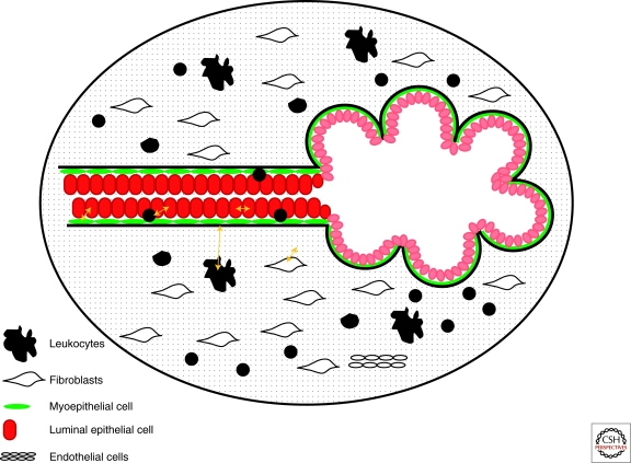
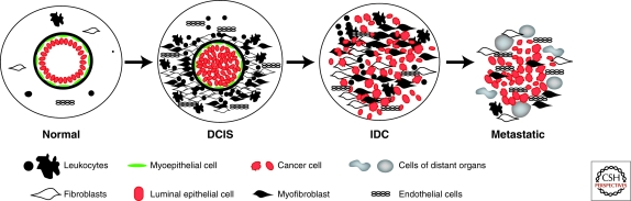
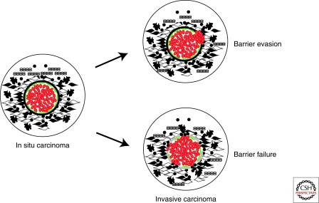
The mammary gland is composed of a diverse array of cell types that form intricate interaction networks essential for its normal development and physiologic function. Abnormalities in these interactions play an important role throughout different stages of tumorigenesis. Branching ducts and alveoli are lined by an inner layer of secretory luminal epithelial cells that produce milk during lactation and are surrounded by contractile myoepithelial cells and basement membrane. The surrounding stroma comprised of extracellular matrix and various cell types including fibroblasts, endothelial cells, and infiltrating leukocytes not only provides a scaffold for the organ, but also regulates mammary epithelial cell function via paracrine, physical, and hormonal interactions. With rare exceptions breast tumors initiate in the epithelial compartment and in their initial phases are confined to the ducts but this barrier brakes down with invasive progression because of a combination of signals emitted by tumor epithelial and various stromal cells. In this article, we overview the importance of cellular interactions and microenvironmental signals in mammary gland development and cancer.


Figures

Figure 1.
Figure 1.
Schematic view of a normal mammary duct and lobule. Black line indicates the basement membrane. In the ducts myoepithelial cells form a nearly complete layer around the luminal epithelial cells, whereas in the alveoli the myoepithelial layer is more fenestrated and some luminal epithelial cells can be in direct contact with the basement membrane. Yellow arrows indicate potential cell–cell and cell–matrix interactions.
Figure 2.
Figure 2.
Microenvironmental alterations during tumor progression. Schematic view of normal breast, DCIS (ductal carcinoma in situ), IDC (invasive ductal carcinoma), and metastatic breast carcinoma. Normal breast ducts are composed of the basement membrane and an inner layer of luminal epithelial sitting on an outer layer of myoepithelial cells. Cells composing the stroma include various leukocytes, fibroblasts, myofibroblasts, and endothelial cells. In DCIS, the myoepithelial cells are epigenetically and phenotypically altered and their number decreases potentially because of signals coming from the tumor epithelial and various stromal cells. The number of stromal fibroblasts, myofibroblasts, lymphocytes, and endothelial cells increases as tumors progress, although the degree of this is tumor subtype-specific.
Figure 3.
Figure 3.
Systemic effects of breast tumors. Tumor cells shed into the circulation and migrate to various organs. At the same time signals emitted by cells within the primary tumor mobilize bone marrow-derived mesenchymal stem cells (BMD-MSCs). These BMD-MSCs are the recruited to the primary tumor and also to other organs such as lungs and brain creating a premetastatic niche prior to the arrival of the disseminated cancer cells. The interaction between cancer cells and BMD-MSCs within the primary tumor enhances the growth, survival, motility, invasive and metastatic capacity of tumor cells via paracrine interactions. In addition, BMD-MSCs can differentiate into myofibroblasts and other stromal cell types that further support the growth and progression of the tumor. Disseminated cancer cells preferentially grow at sites where BMD-MSCs are localized forming distant metastases. Disseminated tumor cells can also return to the original primary tumor and promote its growth and further metastatic spread in a process called “self-seeding.”
Figure 4.
Figure 4.
Hypothetical model depicting two different views of in situ to invasive breast carcinoma transition. Cells are depicted as in Figure 2. In the “barrier evasion” model the tumor epithelial cells disrupt the myoepithelial cell layer, degrade the basement membrane, and migrate into the stroma. In the “barrier failure” model the myoepithelial cells become progressively lost and the basement membrane is disrupted at sites coinciding with areas of leukocytic infiltration and accumulation of myofibroblasts. The loss of the myoepithelial cell layer and basement membrane results in invasive carcinomas where tumor cells can invade surrounding tissues and can migrate to distant organs.

References

    1. Allinen M, Beroukhim R, Cai L, Brennan C, Lahti-Domenici J, Huang H, Porter D, Hu M, Chin L, Richardson A, et al.2004. Molecular characterization of the tumor microenvironment in breast cancer. Cancer Cell 6: 17–32 - PubMed
    1. Anbazhagan R, Osin PP, Bartkova J, Nathan B, Lane EB, Gusterson BA 1998. The development of epithelial phenotypes in the human fetal and infant breast. J Pathol 184: 197–206 - PubMed
    1. Bhowmick NA, Chytil A, Plieth D, Gorska AE, Dumont N, Shappell S, Washington MK, Neilson EG, Moses HL 2004. TGF-β signaling in fibroblasts modulates the oncogenic potential of adjacent epithelia. Science 303: 848–851 - PubMed
    1. Bierie B, Moses HL 2006. Tumour microenvironment: TGF:β The molecular Jekyll and Hyde of cancer. Nat Rev Cancer 6: 506–520 - PubMed
    1. Bierie B, Chung CH, Parker JS, Stover DG, Cheng N, Chytil A, Aakre M, Shyr Y, Moses HL 2009. Abrogation of TGF-β signaling enhances chemokine production and correlates with prognosis in human breast cancer. J Clin Invest 119: 1571–1582 - PMC - PubMed

Publication types

MeSH terms