

Review
doi: 10.1101/cshperspect.a001933.
Epub 2010 Jun 30.
Cellular strategies of axonal pathfinding
Affiliations
- PMID: 20591992
- PMCID: PMC2926747
- DOI: 10.1101/cshperspect.a001933
Item in Clipboard
Review
Cellular strategies of axonal pathfinding
Cold Spring Harb Perspect Biol.
2010 Sep.
Abstract
Axons follow highly stereotyped and reproducible trajectories to their targets. In this review we address the properties of the first pioneer neurons to grow in the developing nervous system and what has been learned over the past several decades about the extracellular and cell surface substrata on which axons grow. We then discuss the types of guidance cues and their receptors that influence axon extension, what determines where cues are expressed, and how axons respond to the cues they encounter in their environment.
Figures
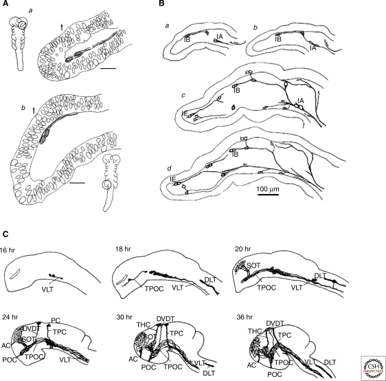
Early axon tract formation in the peripheral and central nervous systems. (A) The axons of the first neurons to differentiate in grasshopper antennae (Aa) and legs (Ab) grow between the surface epithelium and a basement membrane to pioneer axonal pathways from the periphery into the central nervous system. (B a–d) As the limb develops further, progressively more distal neurons differentiate and pioneer short segments of peripheral nerve before fasciculating with more proximal pathways pioneered earlier. (C) In the developing zebrafish CNS, the axons of later differentiating neuronal populations (24–36 h) add onto the earliest axonal pathways (16–20 h), forming a progressively elaborated axonal scaffold over time. (A, Reprinted, with permission, from Bate 1976 [© Nature Publishing Group]; B, reprinted, with permission, from Ho and Goodman 1982 [© Nature Publishing Group]; C, reprinted, with permission, from Ross et al. 1992 [© Society for Neuroscience].)
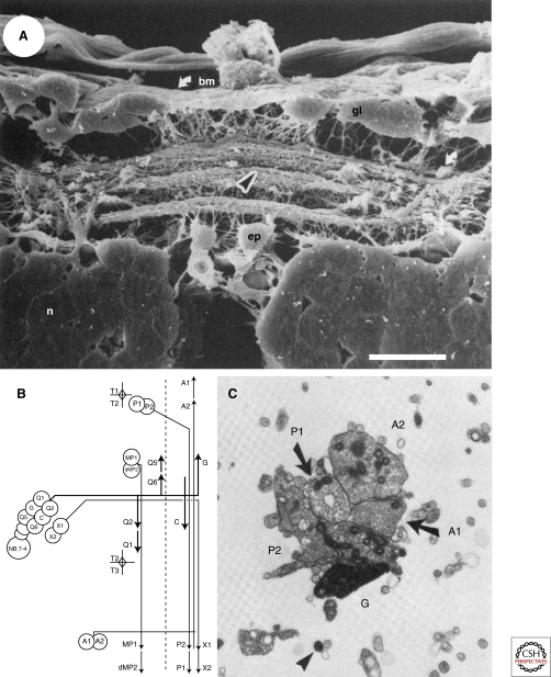
Selective fasciculation in the CNS of the developing grasshopper. (A) Axons are highly fasciculated in the developing grasshopper CNS as visualized by a scanning electron micrograph of the posterior segmental commissure. Specific axons extend within particular bundles. The arrowhead indicates the bundle in which axons growing from the G and C neurons extend. (bm) basement membrane, (gl) glia, (ep) epidermal cells, (n) neurons, scale bar 20 mm. (B) A schematic showing the fasciculation patterns of axons extending from the first neurons born from neuroblast 7–4. The third and fourth born neurons, G and C, extend axons across the midline to a lateral position in the contralateral neuropil and then extend on a specific reproducible axon fascicle that contains the A1, A2, P1, and P2 axons. (C) A transmission electron micrograph of the fascicle containing the G, C, A1, A2, P1, and P2 axons. The G axon was filled with HRP. (A, Reprinted, with permission, from Raper et al. 1983; B, C, reprinted, with permission, from Bastiani et al. 1984 [all © Society for Neuroscience].)
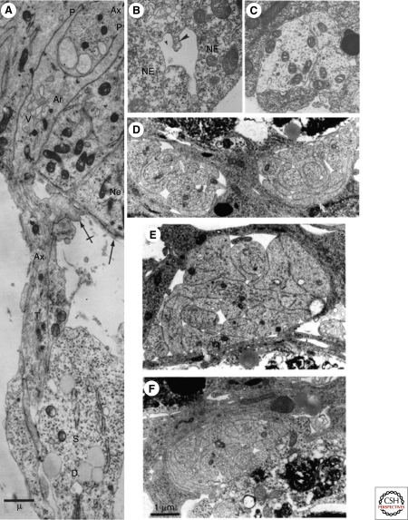
Axons grow in spaces formed by neuroepithelial cells, on other axonal surfaces, and in contact with immature glia. (A) An axonal growth cone enters the neural tube between epithelial cells (Ne) and its tip bifurcates into thin processes (P) that surround a longitudinally extending axon (Ax). A Schwann cell ensheaths the axon but the axon looses this contact before it enters the neural tube. (rabbit, 11–12 days gestation). (B) Spaces or channels form between neuroepithelial cells at the neural tube stage. (C) A few axons course within the spaces formed by neuroepithelial cells (darker cytoplasm) as in B. (Stage 35 and 37, Xenopus). (D–F) E12 mouse embryo, bundle of axons extending in the optic stalk, surrounded by glia (darker cytoplasm). (D) The right bundle contains the flattened lamella of a growth cone of an axon (white arrow) growing adjacent to a glial cell. (E) The axon (white arrowhead) of this growth cone is also apposed to the glial cell; sections in D and E are 200 microns apart. (F) The bottom section is 100 microns more proximal to the retina than the middle section. Two other axons have interposed between the axon in E and the glial wrapping. (A, Reprinted, with permission, from Tennyson 1970 [© Rockefeller University Press]; B, C, reprinted, with permission, from Nordlander and Singer 1982 [© Elsevier]; D–F, reprinted, with permission from Colello and Guillery 1992 [© Wiley].)
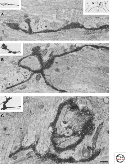
Growth cones contact radial glial processes at the optic chiasm midline. DiI labeled (black deposit) retinal axon growth cones grow in intimate contact with processes of the midline glial palisade (cut in cross section,*) after crossing the midline (A), during pausing (B, inferred from the static image of a complex growth cone), and during a turn away from the midline (C). Coated vesicles are prominent in the glial cells. (Reprinted, with permission, from Marcus et al. 1995 [© Society for Neuroscience].)

Environmental influences on axon pathfinding. Axons navigate through an environment in which neuronal and non-neuronal cells display on their surfaces or secrete into interstitial spaces and the ECM a variety of signaling molecules. These include morphogenic and differentiation factors that influence neuron determination, as well as tropic, modulatory, adhesive, and trophic factors that act directly on the growth cone. Depending on which specific guidance receptors and signaling components are expressed in the growth cone, guidance cues activate particular signaling pathways that regulate growth cone motility. As a result, a growth cone may advance, pause, collapse, withdraw, turn, or fasciculate with other axons.
References
-
- Attardi DG, Sperry RW 1963. Preferential selection of central pathways by regenerating optic fibers. Exp Neurol 7: 46–64 - PubMed
-
- Bagri A, Marín O, Plump AS, Mak J, Pleasure SJ, Rubenstein JL, Tessier-Lavigne M 2002. Slit proteins prevent midline crossing and determine the dorsoventral position of major axonal pathways in the mammalian forebrain. Neuron 33: 233–248 - PubMed
Publication types
MeSH terms
LinkOut - more resources
Full Text Sources
Miscellaneous