. 2010 Sep;84(18):9217-26.
doi: 10.1128/JVI.01069-10. Epub 2010 Jun 30.

Memory CD4 T cells direct protective responses to influenza virus in the lungs through helper-independent mechanisms

Affiliations

Memory CD4 T cells direct protective responses to influenza virus in the lungs through helper-independent mechanisms

John R Teijaro et al. J Virol. 2010 Sep.

Abstract

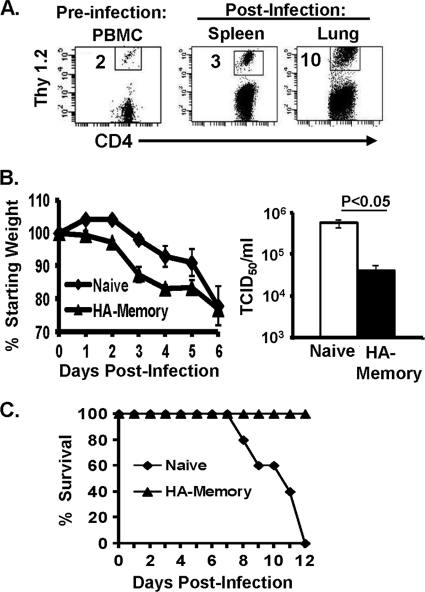
Memory CD4 T cells specific for influenza virus are generated from natural infection and vaccination, persist long-term, and recognize determinants in seasonal and pandemic influenza virus strains. However, the protective potential of these long-lived influenza virus-specific memory CD4 T cells is not clear, including whether CD4 T-cell helper or effector functions are important in secondary antiviral responses. Here we demonstrate that memory CD4 T cells specific for H1N1 influenza virus directed protective responses to influenza virus challenge through intrinsic effector mechanisms, resulting in enhanced viral clearance, recovery from sublethal infection, and full protection from lethal challenge. Mice with influenza virus hemagglutinin (HA)-specific memory CD4 T cells or polyclonal influenza virus-specific memory CD4 T cells exhibited protection from influenza virus challenge that occurred in the presence of CD8-depleting antibodies in B-cell-deficient mice and when CD4 T cells were transferred into lymphocyte-deficient RAG2(-/-) mice. Moreover, the presence of memory CD4 T cells mobilized enhanced T-cell recruitment and immune responses in the lung. Neutralization of gamma interferon (IFN-gamma) production in vivo abrogated memory CD4 T-cell-mediated protection from influenza virus challenge by HA-specific memory T cells and heterosubtypic protection by polyclonal memory CD4 T cells. Our results indicate that memory CD4 T cells can direct enhanced protection from influenza virus infection through mobilization of immune effectors in the lung, independent of their helper functions. These findings have important implications for the generation of universal influenza vaccines by promoting long-lived protective CD4 T-cell responses.


Figures

FIG. 1.
FIG. 1.
Influenza virus-specific memory CD4 T cells mediate secondary responses to influenza virus challenge. (A) Influenza virus infection induces expansion of HA-specific memory CD4 T cells. Flow cytometry plots show the frequencies of CD4+ Thy1.2+ HA-specific memory CD4 T cells in the blood 1 day before infection (left) and in the spleen and lungs 6 days following influenza virus challenge (right), with the percentage of HA-specific Thy1.2+ memory CD4 T cells among total CD4 T cells indicated in each plot. (B) Naive or HA-memory mice were infected intranasally with 500 TCID50 of PR8 influenza virus (sublethal dose) and monitored for 6 days. (Left) Daily weight loss, expressed as a percentage of starting weight (100%), in naive and HA-memory mice following influenza virus challenge (the average weight loss for naive mice was 23%, and that for HA-memory mice was 24%). (Right) Lung viral titers determined by TCID50 assay (see Materials and Methods) of lung homogenates harvested at 6 days postinfection, with a P value of <0.05 for the difference in titers between naive and HA-memory mice (four mice per group). Results are representative of 6 independent experiments. (C) Naive BALB/c or HA-memory mice were infected with a lethal dose of PR8 influenza virus and then monitored daily. The graph shows the percentage of mice surviving each day after infection, with five mice per group. The results are representative of two independent experiments.
FIG. 2.
FIG. 2.
Memory CD4 T-cell-mediated viral clearance in hosts depleted of CD8 T cells. Naive BALB/c and HA-memory mice were left untreated (control) or administered anti-CD8 depleting antibody 1 day prior to and every other day throughout the course of infection. (A) CD8 and CD4 T-cell frequencies in peripheral blood following anti-CD8 depletion in uninfected mice. Plots show CD3ɛ versus CD8β or CD8α in control or anti-CD8-treated mice. (B) Frequencies of CD4 and CD8 T cells in the spleens and lungs of control and CD8-depleted mice 6 days after influenza virus infection. Results are graphed as means ± standard deviations (SD) for five mice per group and are representative of two experiments. Significant differences between control and treated groups were found for CD8 T cells in the spleen (***, P < 0.001) and lungs (**, P < 0.01). (C) Daily weight loss, expressed as a percentage of starting weight (100%), following influenza virus challenge in naive BALB/c or HA-memory mice treated with anti-CD8 depleting antibody or with PBS (control). (D) Lung viral titers determined by TCID50 assay (see Materials and Methods) of lung homogenates harvested at 6 days postinfection. Results were compiled from two independent experiments (n = 8 to 10 mice/group). Significant differences in titers were found between PBS-treated naive and HA-memory mice (*, P < 0.05) and between anti-CD8-treated naive and HA-memory mice (**, P < 0.01).
FIG. 3.
FIG. 3.
Memory CD4 T cells mediate protective responses to influenza virus challenge in the absence of B cells. (A) Influenza virus infection induces expansion of HA-specific memory CD4 T cells in B-cell-deficient (JhD−/−) hosts. Flow cytometry plots show the frequencies of CD4+ Thy1.1+ HA-specific memory CD4 T cells in the blood 1 day before infection (left) and in the spleen and lungs 6 days following influenza virus challenge (right), with the percentage of HA-specific Thy1.1+ memory CD4 T cells among total CD4 T cells indicated in each plot. (B) JhD-memory, JhD naive, and control wild-type naive and HA-memory mice were infected intranasally with 500 TCID50 of PR8 influenza virus and monitored for up to 10 days postinfection. The graph shows daily weights, expressed as percentages of starting weight (100%), for all infected groups. (C) Kinetics of viral clearance in JhD-memory, JhD naive, and control wild-type naive and HA-memory mice at 6 and 10 days postinfection. On day 10, all groups except for JhD naive mice exhibited viral loads below the detection limit of the assay (bd.). Significant differences in titers were found between all groups (P < 0.001) by one-way ANOVA. **, P = 0.005, comparing JhD naive to JhD-memory mice; ***, P = 0.001, comparing naive BALB/c to JhD-memory mice. Results are representative of two independent experiments (7 to 10 mice/group). bd., below detection.
FIG. 4.
FIG. 4.
Protection mediated by polyclonal influenza virus-specific memory CD4 T cells occurs in lymphocyte-deficient hosts. C57BL/6 mice were infected with 500 TCID50 of PR8 influenza virus and monitored for infection, and CD4 T cells were purified from the spleens and lungs of these “flu-memory” mice at 4 to 8 weeks postinfection. (A) Frequencies of influenza virus NP-specific CD4 T cells 8 weeks after infection, as determined by intracellular cytokine staining following stimulation with 1 μg/ml anti-CD28 (medium alone), 1 μg/ml NP311-325 plus 1 μg/ml anti-CD28 (NP311-325), or phorbol myristate acetate-ionomycin (PMA/Iono). These results are representative of two experiments, each with five mice per group. (B) CD4 T cells purified from the spleens and lungs of naive C57BL/6 mice or C57BL/6 flu-memory mice were transferred (8 × 10⁶ total cells, comprising 4 × 10⁶ cells from the spleen and 4 × 10⁶ cells from the lungs) intravenously into RAG2−/− recipients, which were subsequently infected with 500 TCID50 of PR8. The graph shows weight loss morbidities of the influenza virus-infected RAG2−/−, RAG2+Naive, and RAG2+Memory groups up until day 7 postinfection. Weight loss differences between naive and memory groups were not significant. (C) Lung viral titers at day 7 postinfection in RAG2−/−, RAG2+Naive, and RAG2+Memory mice, expressed as the average for 10 mice per group, compiled from two independent experiments with 5 mice per group (representative of three independent experiments). The significance of differences in viral titers between the RAG+Memory, RAG+Naïve, and RAG−/− groups was determined by one-way ANOVA (P = 0.002).
FIG. 5.
FIG. 5.
Localized memory CD4 T-cell-directed responses in the lung are distinct from diffuse T-cell distribution during primary infection. (A) Localization of CD3+ T cells in the lungs of uninfected C57BL/6 mice (top) and influenza virus-infected C57BL/6 mouse recipients of total CD4 T cells from naive (middle left) or C57BL/6 flu-memory (lower left) mice 7 days after infection with X31, using immunohistochemistry. Similar CD3 immunohistochemistry analysis was performed on lungs from PR8-infected RAG2−/− mice that received total CD4 T cells from naive (middle right) or C57BL/6 flu-memory (lower right) mice (see Fig. 6). CD3+ T cells stain brown, as indicated by arrows, and images for representative animals of five mice per group are shown at a magnification of ×20. (B) CD3 T cells were enumerated by counting the total number of cells staining for CD3 and taking the average for five 1-mm2 sections of lung per animal. Each section counted had a similar lung architecture. Data shown are the average counts for five animals per group. ***, P < 0.001, representing the difference in CD3 numbers between groups, as determined by ANOVA.
FIG. 6.
FIG. 6.
Memory CD4 T-cell-mediated protection is dependent on IFN-γ. Naive and HA-memory mice were treated intraperitoneally and intranasally with control IgG and anti-IFN-γ antibody (see Materials and Methods) and then challenged with 500 TCID50 of PR8 influenza virus. (A) Daily weight loss, measured as described previously. (B) Lung viral titers at 5 days post-influenza virus challenge, determined by the TCID50 method as described previously. Results are representative of 2 independent experiments (8 to 10 mice/group). *, P < 0.05, comparing HA-memory mice treated with XMG1.2 and IgG1. (C) Absolute numbers of HA-specific memory CD4 T cells in spleen and lung tissue, calculated from flow cytometry analysis and microscopic cell counts. Data are shown as the total number of Thy1.2+ memory CD4 T cells in each tissue. (D) IFN-γ neutralization does not skew cytokine production of HA-memory CD4 T cells after influenza virus infection. HA-specific memory CD4 T cells were isolated at 5 days postinfection from the spleen and lungs of influenza virus-infected HA-memory mice and stimulated ex vivo with PMA-ionomycin for 5 h. The graphs show percentages of Thy1.2+ memory CD4 T cells producing IFN-γ, IL-4, and IL-17 in both spleen and lung tissues.
FIG. 7.
FIG. 7.
Heterosubtypic protection mediated by memory CD4 T cells requires IFN-γ secretion. CD4 T cells containing influenza virus-specific memory CD4 T cells were purified from the spleens and lungs of C57BL/6 mice infected with 500 TCID50 of PR8 influenza virus 6 weeks previously, as described in the legend to Fig. 5A, and 8 × 10⁶ CD4 T cells were transferred into B6.Ly5.2 congenic hosts. The resultant hosts of polyclonal memory CD4 T cells were challenged with 10,000 TCID50 of the heterosubtypic strain HK-X31 and then administered control IgG or anti-IFN-γ (XMG1.2) antibody at day 0 and every other day throughout infection. (A) Weight losses of differentially treated mouse memory recipients were plotted as percentages of starting weights. (B) Viral titers were determined by MDCK assay at 7 days postinfection and are shown as averages for five mice per group, with the P value of <0.001 representing a difference in titers between memory mice treated with either control antibody or anti-IFN-γ antibody. The results are representative of three independent experiments. (C) Lung histology was determined by hematoxylin and eosin staining at 7 days postinfection. An image for a representative animal is shown for each group (five mice per group) and for a lung from an uninfected animal.

References

    1. Ahmadzadeh, M., and D. L. Farber. 2002. Functional plasticity of an antigen-specific memory CD4 T cell population. Proc. Natl. Acad. Sci. U. S. A. 99:11802-11807. - PMC - PubMed
    1. Allan, W., Z. Tabi, A. Cleary, and P. C. Doherty. 1990. Cellular events in the lymph node and lung of mice with influenza. Consequences of depleting CD4+ T cells. J. Immunol. 144:3980-3986. - PubMed
    1. Assarsson, E., H. H. Bui, J. Sidney, Q. Zhang, J. Glenn, C. Oseroff, I. N. Mbawuike, J. Alexander, M. J. Newman, H. Grey, and A. Sette. 2008. Immunomic analysis of the repertoire of T-cell specificities for influenza A virus in humans. J. Virol. 82:12241-12251. - PMC - PubMed
    1. Belz, G. T., D. Wodarz, G. Diaz, M. A. Nowak, and P. C. Doherty. 2002. Compromised influenza virus-specific CD8(+)-T-cell memory in CD4(+)-T-cell-deficient mice. J. Virol. 76:12388-12393. - PMC - PubMed
    1. Bender, B. S., T. Croghan, L. Zhang, and P. A. Small, Jr. 1992. Transgenic mice lacking class I major histocompatibility complex-restricted T cells have delayed viral clearance and increased mortality after influenza virus challenge. J. Exp. Med. 175:1143-1145. - PMC - PubMed

Publication types

MeSH terms

Substances