. 2010 Sep;84(18):9267-77.
doi: 10.1128/JVI.00616-10. Epub 2010 Jun 30.

All three domains of the hepatitis C virus nonstructural NS5A protein contribute to RNA binding

Affiliations

All three domains of the hepatitis C virus nonstructural NS5A protein contribute to RNA binding

Toshana L Foster et al. J Virol. 2010 Sep.

Abstract

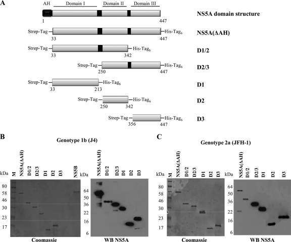
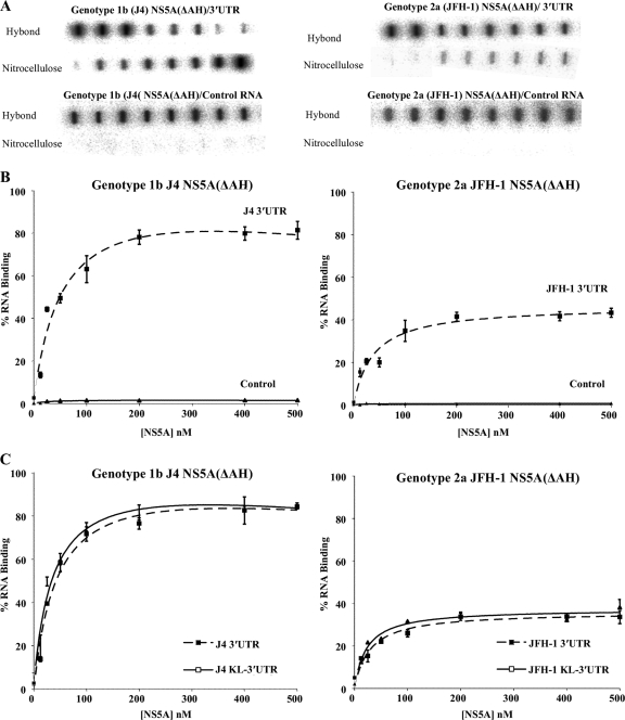
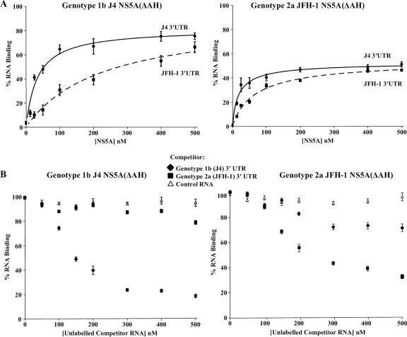
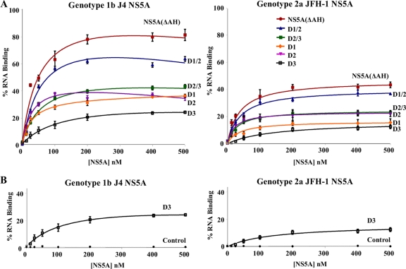
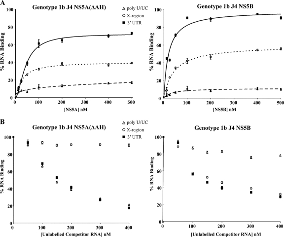
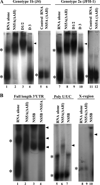
The hepatitis C virus (HCV) nonstructural protein NS5A is critical for viral genome replication and is thought to interact directly with both the RNA-dependent RNA polymerase, NS5B, and viral RNA. NS5A consists of three domains which have, as yet, undefined roles in viral replication and assembly. In order to define the regions that mediate the interaction with RNA, specifically the HCV 3' untranslated region (UTR) positive-strand RNA, constructs of different domain combinations were cloned, bacterially expressed, and purified to homogeneity. Each of these purified proteins was probed for its ability to interact with the 3' UTR RNA using filter binding and gel electrophoretic mobility shift assays, revealing differences in their RNA binding efficiencies and affinities. A specific interaction between domains I and II of NS5A and the 3' UTR RNA was identified, suggesting that these are the RNA binding domains of NS5A. Domain III showed low in vitro RNA binding capacity. Filter binding and competition analyses identified differences between NS5A and NS5B in their specificities for defined regions of the 3' UTR. The preference of NS5A, in contrast to NS5B, for the polypyrimidine tract highlights an aspect of 3' UTR RNA recognition by NS5A which may play a role in the control or enhancement of HCV genome replication.


Figures

FIG. 1.
FIG. 1.
The HCV 3′ UTR RNA. (A) The positive-strand 3′ UTR consists of three distinct regions, i.e., a short genotype-specific variable region, a polypyrimidine tract [poly(U/UC)] of variable length, and a conserved 98-nucleotide sequence known as the X region containing three stable stem-loops. The predicted structure of the genotype 1b 3′ UTR is shown. (B) Left panel, the integrities of in vitro-transcribed radiolabeled full-length 3′ UTR RNAs of genotypes 1b (nucleotides 9375 to 9595) and 2a (nucleotides 9443 to 9678) and the poly(U/UC) (nucleotides 9406 to 9497) and X region (nucleotides 9498 to 9595) of genotype 1b are shown on denaturing polyacrylamide gels. Right panel, the integrities of in vitro-transcribed radiolabeled RNAs comprising the 3′-terminal NS5B-coding region plus the 3′ UTR RNAs of genotypes 1b (nucleotides 9136 to 9595) and 2a (nucleotides 9204 to 9678) (KL-3′ UTR) are shown on denaturing polyacrylamide gels.
FIG. 2.
FIG. 2.
Domain structure and expression of HCV NS5A. (A) Schematic diagram of the functional domains of NS5A and design of the constructs used in the study (genotype 1b NS5A protein numbering). The N-terminal amphipathic helix of NS5A (black box) is responsible for the interaction of NS5A with membranes. NS5A is organized into three domains that are separated by low-complexity sequences, indicated by black boxes. The NS5A constructs used all lacked the N-terminal amphipathic helix and were designed to include an N-terminal Strep tag and a C-terminal hexahistidine tag. (B and C) SDS-PAGE and Western blot analysis of the NS5A(ΔAH) and NS5A domain constructs purified by nickel affinity and Streptactin tag affinity chromatography. Coomassie brilliant blue-stained gels and Western blots (WB) using anti-NS5A antibodies for NS5A proteins of genotype 1b strain J4 (B) and genotype 2a strain JFH-1 (C) are shown.
FIG. 3.
FIG. 3.
Filter binding analysis of the interaction between NS5A(ΔAH) and the HCV 3′ UTR RNA. (A) The indicated proteins were incubated with radiolabeled RNA (1 nM), either 3′ UTR (upper panel) or control FMDV 3C aptamer (lower panel), before application to a slot blot apparatus, filtering through nitrocellulose and Hybond-N membranes, and visualization by phosphorimaging. From left to right, the slots contained 0, 12.5, 25, 50, 100, 200, 400, and 500 nM protein. (B) The percentage of RNA bound to the nitrocellulose membrane was quantified and plotted as a function of the NS5A concentration. The data were fitted to a hyperbolic equation. Experiments were performed in triplicate, and the means and standard errors are plotted. (C) NS5A(ΔAH) (0 to 500 nM) was incubated with the corresponding genotype radiolabeled RNA (1 nM), corresponding to either the 3′ UTR or the 3′ terminal NS5B coding region plus 3′ UTR (KL-3′ UTR). Experiments were performed in triplicate, and the means and standard errors are plotted.
FIG. 4.
FIG. 4.
Filter binding analysis of the genotype specificity of the NS5A-3′ UTR interaction. (A) Purified NS5A(ΔAH) of genotype 1b (J4, left) or genotype 2a (JFH-1, right) was incubated with radiolabeled 3′ UTR RNA (1 nM) or either genotype 1b (J4, solid lines) or genotype 2a (JFH-1, dashed lines) and analyzed as described for Fig. 3. The percentage of RNA bound to the nitrocellulose membrane was quantified and plotted as a function of the NS5A concentration. The data were fitted to a hyperbolic equation. (B) Purified NS5A(ΔAH) of genotype 1b (J4, left) or genotype 2a (JFH-1, right) was incubated with 1 nM radiolabeled 3′ UTR RNA in the presence of increasing concentrations (0 to 500 nM) of unlabeled 3′ UTR of either genotype 1b or 2a or control FMDV 3C aptamer RNA. The percentage of RNA bound to the nitrocellulose membrane was quantified and plotted as a function of the competitor RNA concentration.
FIG. 5.
FIG. 5.
The three domains of NS5A exhibit different RNA binding properties in vitro. (A) Purified NS5A(ΔAH) and either combinations of domains or individual domains of genotype 1b (J4, left) or genotype 2a (JFH-1, right) were incubated with radiolabeled 3′ UTR RNA (1 nM) of the corresponding genotype and analyzed as described for Fig. 3. The percentage of RNA bound to the nitrocellulose membrane was quantified and plotted as a function of the NS5A concentration. The data were fitted to a hyperbolic equation. (B) Purified D3 (domain III alone) of genotype 1b (J4, left) or genotype 2a (JFH-1, right) was incubated with radiolabeled 3′ UTR RNA (1 nM) of the corresponding genotype or a control FMDV 3C aptamer RNA and analyzed as described for Fig. 3. In all cases experiments were performed in triplicate, and the means and standard errors are plotted.
FIG. 6.
FIG. 6.
Binding specificity of the in vitro 3′ UTR-NS5A interaction. (A) Purified NS5A(ΔAH) (left) or NS5B (right) of genotype 1b was incubated with radiolabeled 3′ UTR RNA (solid lines), poly(U/UC) (dashed lines), or X region RNA (dotted lines) (all at 1 nM) and analyzed as described for Fig. 3. The percentage of RNA bound to the nitrocellulose membrane was quantified and plotted as a function of the NS5A concentration. The data were fitted to a hyperbolic equation. Experiments were performed in triplicate, and the means and standard errors are plotted. (B) NS5A(ΔAH) (left) and NS5B (right) proteins were bound to 1 nM radiolabeled 3′ UTR in the presence of increasing concentrations (0 to 400 nM) unlabeled 3′ UTR, poly(U/UC), or X region RNA.
FIG. 7.
FIG. 7.
Electrophoretic mobility shift analysis of NS5A-3′ UTR RNA binding. (A) Radiolabeled 3′ UTR RNA or control FMDV 3C aptamer was incubated with 200 nM NS5A(ΔAH), D1/2, or D3 proteins as indicated, prior to separation by native acrylamide gel electrophoresis and visualization by autoradiography. The positions of the major retarded NS5A-RNA complexes are indicated by black triangles, and the asterisks mark the positions of the unbound 3′ UTR RNA (the two bands most likely represent alternative conformations of the RNA). (B) Radiolabeled RNA corresponding to the 3′ UTR, poly(U/UC), or X region was incubated with 100 nM NS5A(ΔAH) or NS5B or a combination. The positions of the major retarded NS5A-RNA and NS5B-RNA complexes are indicated by black triangles, and the asterisks mark the positions of the unbound 3′ UTR RNA.

Similar articles

Cited by

References

    1. Appel, N., M. Zayas, S. Miller, J. Krijnse-Locker, T. Schaller, P. Friebe, S. Kallis, U. Engel, and R. Bartenschlager. 2008. Essential role of domain III of nonstructural protein 5A for hepatitis C virus infectious particle assembly. PLoS Pathog. 4:e1000035. - PMC - PubMed
    1. Cheng, J., C. Ming-Fu, and S. C. Chang. 1999. Specific interaction between the hepatitis C virus NS5B RNA polymerase and the 3′ end of the viral RNA. J. Virol. 73:7044-7049. - PMC - PubMed
    1. Choo, Q. L., K. H. Richman, J. H. Han, K. Berger, C. Lee, C. Dong, C. Gallegos, D. Coit, R. Medina-Selby, and P. J. Barr. 1991. Genetic organization and diversity of the hepatitis C virus. Proc. Natl. Acad. Sci. U. S. A. 88:2451-2455. - PMC - PubMed
    1. Foster, G., and P. Mathurin. 2008. Hepatitis C virus therapy to date. Antivir. Ther. 13:1-8. - PubMed
    1. Friebe, P., J. Boudet, J.-P. Simorre, and R. Bartenschlager. 2005. Kissing-loop interaction in the 3′ end of the hepatitis C virus genome essential for RNA replication. J. Virol. 79:380-392. - PMC - PubMed

Publication types

Substances

LinkOut - more resources