. 2010 Sep;84(18):9047-58.
doi: 10.1128/JVI.00698-10. Epub 2010 Jun 30.

Human enterovirus 109: a novel interspecies recombinant enterovirus isolated from a case of acute pediatric respiratory illness in Nicaragua

Affiliations

Human enterovirus 109: a novel interspecies recombinant enterovirus isolated from a case of acute pediatric respiratory illness in Nicaragua

Nathan L Yozwiak et al. J Virol. 2010 Sep.

Abstract

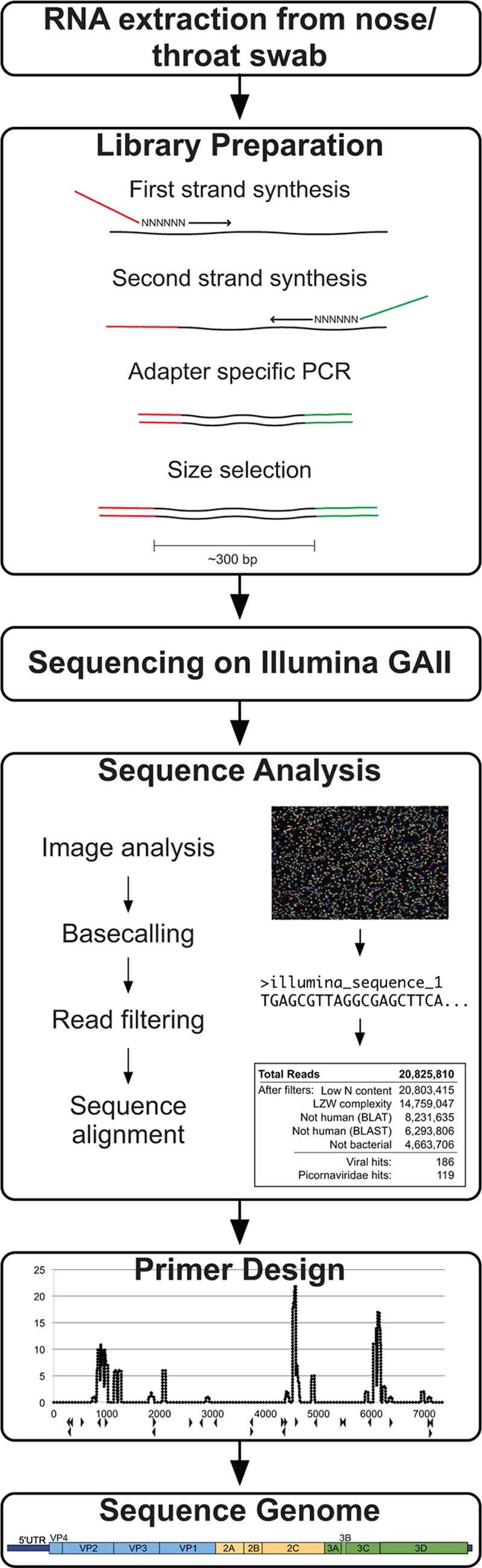
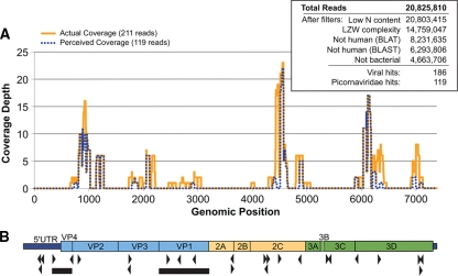
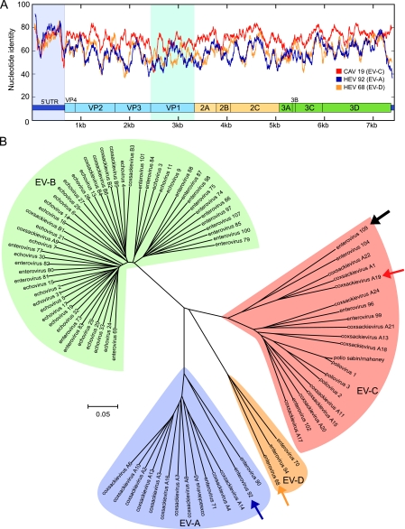
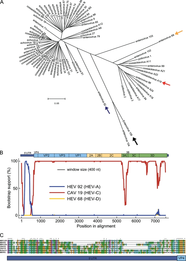
Enteroviruses (Picornaviridae family) are a common cause of human illness worldwide and are associated with diverse clinical syndromes, including asymptomatic infection, respiratory illness, gastroenteritis, and meningitis. In this study, we report the identification and complete genome sequence of a novel enterovirus isolated from a case of acute respiratory illness in a Nicaraguan child. Unbiased deep sequencing of nucleic acids from a nose and throat swab sample enabled rapid recovery of the full-genome sequence. Phylogenetic analysis revealed that human enterovirus 109 (EV109) is most closely related to serotypes of human enterovirus species C (HEV-C) in all genomic regions except the 5' untranslated region (5' UTR). Bootstrap analysis indicates that the 5' UTR of EV109 is likely the product of an interspecies recombination event between ancestral members of the HEV-A and HEV-C groups. Overall, the EV109 coding region shares 67 to 72% nucleotide sequence identity with its nearest relatives. EV109 isolates were detected in 5/310 (1.6%) of nose and throat swab samples collected from children in a pediatric cohort study of influenza-like illness in Managua, Nicaragua, between June 2007 and June 2008. Further experimentation is required to more fully characterize the pathogenic role, disease associations, and global distribution of EV109.


Figures

FIG. 1.
FIG. 1.
Viral recovery workflow. The method for viral genome recovery begins with RNA extraction from a nose/throat swab of clinical interest. A sequencing library is generated from the nucleic acids using reverse transcription and random-primed amplification to affix known sequencing adapter molecules necessary for the Illumina platform. The Illumina GAII sequencing is performed using the manufacturer's protocol. Sequence analysis includes image analysis and base-calling software, read filtering using quality metrics, and iterative sequence alignments. Reads with similarity to the viral sequence of interest are computationally assembled or used for primer design for subsequent specific amplification of remaining viral regions.
FIG. 2.
FIG. 2.
Deep-sequencing results and picornavirus genome coverage. (A) Coverage map of deep-sequencing-derived picornavirus reads and their adjusted EV109 genome positions before (perceived coverage) and after (actual coverage) full-genome sequencing. (Inset) Summary of total 61-nt deep-sequencing reads and results from the iterative alignment analysis. (B) Schematic diagram of EV109 genome. The black triangles denote primer positions derived from deep-sequencing reads used to recover the full-length EV109 genome. The black bars denote sequence regions recovered from all five EV109 isolates. The predicted EV109 genome domains are drawn to scale.
FIG. 3.
FIG. 3.
Relationships between known enteroviruses and EV109 based on full-length genome analysis. (A) Full-genome similarity plot depicting scanning pairwise identity using a 100-nt sliding window evaluated at each nucleotide. The EV109 sequence is compared with a close HEV-C relative, coxsackievirus A19 (CAV19) and more distant HEV-A (EV92) and HEV-D (EV68) serotypes. The conserved enteroviral domains are depicted to scale. The 5′ UTR and VP1 regions are highlighted. (B) Phylogenetic tree constructed from complete enterovirus genomes. The EV109 genome sequenced in this study is depicted with a solid black arrow, coxsackievirus A19 is identified by a red arrow, enterovirus 92 is identified by a purple arrow, and enterovirus 68 is identified by a yellow arrow. ClustalW and MEGA were used for alignments and tree construction, respectively, using the neighbor-joining method and 500 bootstrap replicates.
FIG. 4.
FIG. 4.
Sequence analysis and RNA folding predictions reveal conserved picornavirus features in EV109. RNA secondary structure prediction of the EV109 5′ UTR using pknotsRG (35) and visualized using PseudoViewer, version 3 (7). Previously described type 1 IRES features maintained in EV109 include a 5′ cloverleaf as stem-loop I, a GNRA sequence in stem-loop IV, and a pyrimidine-rich region in stem-loop VI (29). Stem-loops II and III are predicted as one combined stem-loop structure. Other nucleic acid folding software (Mfold and nupack) predicted similar secondary structures (10, 51).
FIG. 5.
FIG. 5.
The EV109 5′ UTR region reveals evidence of an ancestral recombination event. (A) Phylogenetic tree constructed from 5′ UTR regions of 74 annotated human enteroviruses. A black arrow denotes EV109. Also highlighted are CAV19 (red arrow), EV92 (blue arrow), and EV68 (yellow arrow), which are used in the subsequent bootscanning analysis. ClustalW and MEGA were used for alignments and tree construction, respectively, using the neighbor-joining method and 500 bootstrap replicates. (B) Bootscanning analysis of EV109. Bootscanning analysis was performed with other serotypes of HEV-A (EV92), HEV-C (CAV19), and HEV-D (EV68) using a word size of 400 and step size of 20. (C) Sequence alignment depicting the 5′ UTR-VP4 junction site. The EV109 sequence is compared to representative members of HEV-A and HEV-C.
FIG. 6.
FIG. 6.
Amino acid conservation on the enterovirus capsid pentamer subunit. Amino acid alignment PSSM score of EV109 compared to other HEV-C capsid sequences and mapped on the pentamer crystal structure of coxsackievirus A21. Residues shaded in blue have a higher PSSM score and are more conserved in EV109, whereas yellow residues have a negative PSSM and are nonconserved. A scale bar is given below panel C. (A) External pentamer view. (B) Cross-sectional view. (C) Internal pentamer view.

Similar articles

Cited by

References

    1. Aminev, A. G., S. P. Amineva, and A. C. Palmenberg. 2003. Encephalomyocarditis virus (EMCV) proteins 2A and 3BCD localize to nuclei and inhibit cellular mRNA transcription but not rRNA transcription. Virus Res. 95:59-73. - PubMed
    1. Amineva, S. P., A. G. Aminev, A. C. Palmenberg, and J. E. Gern. 2004. Rhinovirus 3C protease precursors 3CD and 3CD′ localize to the nuclei of infected cells. J. Gen. Virol. 85:2969-2979. - PubMed
    1. Andersson, P., K. Edman, and A. M. Lindberg. 2002. Molecular analysis of the echovirus 18 prototype: evidence of interserotypic recombination with echovirus 9. Virus Res. 85:71-83. - PubMed
    1. Bazan, J. F., and R. J. Fletterick. 1988. Viral cysteine proteases are homologous to the trypsin-like family of serine proteases: structural and functional implications. Proc. Natl. Acad. Sci. U. S. A. 85:7872-7876. - PMC - PubMed
    1. Bolanaki, E., C. Kottaridi, P. Markoulatos, L. Margaritis, and T. Katsorchis. 2005. A comparative amplification of five different genomic regions on Coxsackie A and B viruses. Implications in clinical diagnostics. Mol. Cell. Probes 19:127-135. - PubMed

Publication types

LinkOut - more resources