. 2010 Sep;84(18):9516-32.
doi: 10.1128/JVI.00775-10. Epub 2010 Jun 30.

A short N-terminal peptide motif on flavivirus nonstructural protein NS1 modulates cellular targeting and immune recognition

Affiliations

A short N-terminal peptide motif on flavivirus nonstructural protein NS1 modulates cellular targeting and immune recognition

Soonjeon Youn et al. J Virol. 2010 Sep.

Abstract

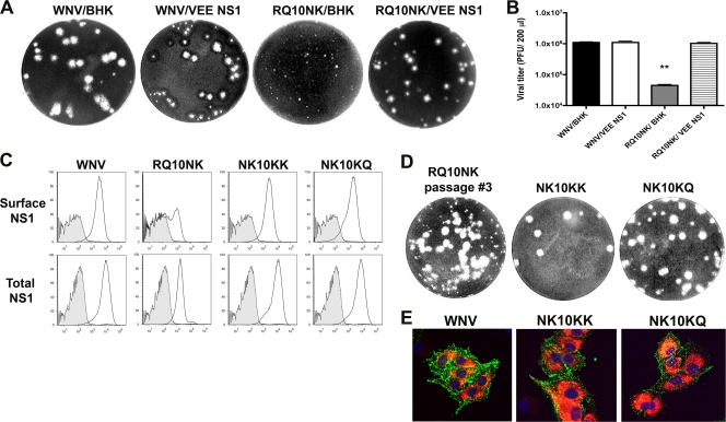
Flavivirus NS1 is a versatile nonstructural glycoprotein, with intracellular NS1 functioning as an essential cofactor for viral replication and cell surface and secreted NS1 antagonizing complement activation. Even though NS1 has multiple functions that contribute to virulence, the genetic determinants that regulate the spatial distribution of NS1 in cells among different flaviviruses remain uncharacterized. Here, by creating a panel of West Nile virus-dengue virus (WNV-DENV) NS1 chimeras and site-specific mutants, we identified a novel, short peptide motif immediately C-terminal to the signal sequence cleavage position that regulates its transit time through the endoplasmic reticulum and differentially directs NS1 for secretion or plasma membrane expression. Exchange of two amino acids within this motif reciprocally changed the cellular targeting pattern of DENV or WNV NS1. For WNV, this substitution also modulated infectivity and antibody-induced phagocytosis of infected cells. Analysis of a mutant lacking all three conserved N-linked glycosylation sites revealed an independent requirement of N-linked glycans for secretion but not for plasma membrane expression of WNV NS1. Collectively, our experiments define the requirements for cellular targeting of NS1, with implications for the protective host responses, immune antagonism, and association with the host cell sorting machinery. These studies also suggest a link between the effects of NS1 on viral replication and the levels of secreted or cell surface NS1.


Figures

FIG. 1.
FIG. 1.
Differential staining of NS1 on DENV- and WNV-infected cells. (A) Confocal microscopy. BHK21 cells were mock infected or infected with DENV (MOI of 1) or WNV (MOI of 0.1) and harvested 24 h later. A higher MOI was used for DENV to obtain the same relative level of infection as it replicates more slowly. Cells were cooled on ice and stained for NS1 on the surface using 2G6 (DENV) or 17NS1 (WNV) mouse MAbs. Subsequently, cells were fixed, permeabilized, and then stained for intracellular E protein using the cross-reactive humanized WNV E18 MAb. Nuclei were counterstained with TO-PRO-3 (blue). The images were analyzed using a LSM 510 confocal microscope. Scale bar, 20 μm. (B) Flow cytometry. BHK21 cells were infected with DENV or WNV as detailed above. Total NS1 or E was determined after cell permeabilization and indirect immunofluorescence staining with the cross-reactive MAb 9NS1 or WNV E18. Surface levels of NS1 were assessed by staining live cells at 4°C with the cross-reactive MAb 9NS1. The y axis indicates the number of cells, and the x axis shows relative NS1 or E levels. Shaded histograms are isotype controls. For microscopic and flow cytometric analyses, data are representative of at least three independent experiments.
FIG. 2.
FIG. 2.
NS1 on the surface of flavivirus-infected cells is targeted directly to the plasma membrane. (A) Sodium chlorate inhibits binding of soluble NS1 to uninfected cells. Uninfected BHK21 cells were pretreated with medium or medium supplemented with 10 mM sodium chlorate, which inhibits the surface expression of glycosaminoglycans (5), and then incubated with soluble WNV NS1 for 2 h at 4°C. Cells were washed extensively and stained for NS1 on the surface using the 9NS1 MAb. (B and C) Sodium chlorate does not inhibit surface expression of NS1 on WNV-infected cells. BHK21 cells were infected with WNV (MOI of 0.1) or treated with medium or medium supplemented with 10 mM sodium chlorate, and 1 day later live cells were immunostained on ice with 9NS1 (B) or 4NS1 (C) MAb to determine the levels of surface NS1 by confocal microscopy (B) or flow cytometry (C). Subsequently, for panel B the same cells were fixed, permeabilized, and stained with chimeric human 17NS1 MAb to confirm the total levels of NS1. For the flow cytometry data, the y axis indicates the number of cells, and the x axis shows relative NS1 levels. Shaded histograms are isotype controls. The data are representative of two independent experiments.
FIG. 3.
FIG. 3.
Expression of DENV and WNV NS1 in BHK21 cells using a recombinant infectious SINV. (A) Scheme of transgenic expression of NS1. Full-length DENV or WNV NS1 was cloned downstream of a second subgenomic 26S promoter as indicated in the cartoon. Transfection of in vitro derived RNA produces infectious SINV that encodes a flavivirus NS1 transgene. (B) Confocal microscopy. BHK21 cells were mock infected or infected with SINV-DENV-NS1, SINV-WNV-NS1, or SINV-WNV-NS1-NS2A1-24 (MOI of 0.1) and harvested at 14 h after infection. Live cells were cooled to 4°C and stained for NS1 on the surface using the cross-reactive 9NS1 MAb. Nuclei were counterstained with TO-PRO-3. A lower MOI was used in the microscopic experiments to facilitate visualization of individual infected cells. (C) Flow cytometry. BHK21 cells were infected with SINV-DENV-NS1 or SINV-WNV-NS1 at an MOI of 1 as detailed above. Total DENV NS1, WNV NS1, and SINV envelope proteins were determined after cell permeabilization and indirect immunofluorescence staining with the cross-reactive MAb 9NS1 or anti-SINV mouse ascites fluid. Surface NS1 levels were assessed by staining live intact cells at 4°C. The y axis indicates the number of cells, and the x axis shows relative NS1 and E2 levels. Shaded histograms are isotype controls. The data are representative of at least three independent experiments.
FIG. 4.
FIG. 4.
The effect of N-linked glycosylation on cell surface expression of NS1 using a recombinant infectious WNV or transgenic SINV. A glycosylation null mutant of WNV NS1 (CHO null) was generated by iterative site-directed mutagenesis in a shuttle vector and then cloned into the infectious cDNA of a New York 1999 strain (385-99) of WNV. (A) BHK21 cells were infected with WNV expressing wild-type NS1 (MOI of 0.1) or NS1 CHO null (MOI of 3) and subjected to Western blotting with 8NS1 MAb under reducing conditions. A higher MOI was required for the CHO null WNV because of a growth defect of this virus, as reported previously (62). The difference in NS1 size of the CHO null mutant is due to the absence of N-linked glycans. Both wild-type and mutant NS1s have additional higher-molecular-weight NS1′ forms, which are indicated and are due to ribosomal frameshifting (25, 47). (B) Confocal microscopy. BHK21 cells were infected with wild-type or NS1 CHO null WNV and harvested at 24 h after infection. Live cells were cooled to 4°C and stained for NS1 on the surface using the cross-reactive 9NS1 MAb (green). Subsequently, cells were fixed and permeabilized and stained with chimeric human 17NS1 MAb to confirm expression of NS1 (red). Nuclei were counterstained with TO-PRO-3. (C) Flow cytometry. BHK21 cells were infected with SINV-WNV-NS1 CHO null or SINV-WNV-NS1 as detailed in the legend of Fig. 2 and harvested at 14 h after infection. Total WNV NS1 or SINV E2 was determined after cell fixation, permeabilization, and indirect immunofluorescence staining. Surface NS1 levels were assessed by staining live intact cells at 4°C. The y axis indicates the number of cells, and the x axis shows relative NS1 levels. Shaded histograms are isotype controls. The data are representative of at least three independent experiments.
FIG. 5.
FIG. 5.
Scheme and phenotypes of the WNV-DENV NS1 chimeras. (A) The construction and expression of NS1 chimeras are detailed in Materials and Methods. The most N-terminal box indicates the origin of the signal peptide (black, WNV NS1; red, DENV NS1). The remaining boxes denote the regions that are comprised of WNV NS1 (blue) or DENV NS1 (white). The numbers correspond to the amino acid position of NS1 at the breakpoint of the chimera. Encircled plus signs denote encoded N-linked glycosylation sites; wild-type DENV and WNV NS1s have two and three N-linked glycans, respectively. The surface expression phenotype for each NS1 variant, when expressed in a recombinant SINV, is listed immediately to the right of the chimera: W and D indicate a WNV-like or DENV-like surface pattern, respectively. The analysis is based on at least three independent experiments with each chimera. (B) Confocal microscopy. BHK21 cells were infected with SINV-WNV-NS1 (WNV NS1), SINV-DENV NS1 (DENV NS1), or the indicated chimera (MOI of 0.1) and harvested at 14 h after infection. Live cells were cooled to 4°C and stained for NS1 on the surface using the cross-reactive 9NS1 MAb. Nuclei were counterstained with TO-PRO-3. The data are representative of two or three independent experiments.
FIG. 6.
FIG. 6.
Phenotype of site-specific variants in the N-terminal region of NS1. (A) Sequence alignment of flavivirus NS1 immediately C-terminal to the signal peptide cleavage site. The numbers above correspond to the amino acid sequence of NS1. The sequences highlighted in green and yellow comprise a region of dissimilarity between the two viruses. Residue 10 shows significant variation between all serotypes of DENV and other flaviviruses. (B) Confocal microscopy. BHK21 cells were infected with SINV-WNV-NS1 (WT) or a series of mutants (AI5VV, ID6VS, IS8WK, RQ10NK, and R10N) that incorporated the corresponding residues of DENV NS1 and harvested at 14 h after infection. Cells were cooled to 4°C and stained for NS1 on the surface using the 4NS1 MAb (green). Subsequently, cells were fixed and permeabilized and stained with chimeric human 17NS1 MAb to confirm expression of NS1 (red). Nuclei were counterstained with TO-PRO-3. The data are representative of two to three independent experiments. (C) Flow cytometric analysis. BHK21 cells were infected with SINV-WNV-NS1 or the indicated mutants as detailed above. Staining of live intact cells at 4°C assessed the surface levels of wild-type and variant NS1 using the 4NS1 MAb. Total NS1 and SINV E2 were determined after fixation, cell permeabilization, and immunofluorescence staining with anti-NS1 or anti-SINV E2 antibodies. The y axis indicates the number of cells, and the x axis shows the relative NS1 or E2 level. Shaded histograms are isotype controls. Data are representative of at least three independent experiments.
FIG. 7.
FIG. 7.
Phenotype of site-specific variants in the N-terminal region of DENV-2 NS1. (A) Flow cytometric analysis. BHK21 cells were infected with SINV-DENV-2 wild-type (WT) NS1 or the mutant SINV-DENV-2 NK10RQ that incorporated the corresponding residues of WNV NS1. Staining of live intact cells at 4°C assessed the surface levels of wild-type and variant NS1 using the cross-reactive 9NS1 MAb. Total DENV NS1 and SINV E2 were determined after fixation, cell permeabilization, and immunofluorescence staining with anti-NS1 or anti-SINV E2 antibodies. The y axis indicates the number of cells, and the x axis shows relative DENV NS1 or SINV E2 level. Shaded histograms are isotype controls. Data are representative of at least two independent experiments. (B) Confocal microscopy. BHK21 cells were infected with SINV-DENV-2 WT NS1 or the mutant SINV-DENV-2 NK10RQ and harvested at 14 h after infection. Cells were cooled to 4°C and stained for NS1 on the surface using the 9NS1 MAb (green). Nuclei were counterstained with TO-PRO-3. The data are representative of two independent experiments. (C) Western blotting with the 1F11 anti-DENV-2 NS1 MAb of supernatants and cell lysates from BHK21 cells infected with SINV-DENV-2 WT NS1 or SINV-DENV-2 NK10RQ. Note that the levels of NS1 in the supernatant of the SINV-DENV-2-NK10RQ samples are lower and that those in the cell lysate are slightly higher.
FIG. 8.
FIG. 8.
Biosynthesis of wild-type and nutant WNV NS1. (A) BHK21 cells were infected with different SINV-NS1 for 12 h, placed in cysteine-methionine-free medium for 20 min, pulsed with [35S]cysteine-methionine for 20 min, and then chased for 1 or 6 h. Cell supernatants or lysates were harvested, immunoprecipitated with 9NS1 MAb-Sepharose, and digested with endo H (H) or PNGase F (F). (B) A more detailed early 35S pulse-chase time course was performed with wild-type NS1, IS8WK, and RQ10NK with recombinant SINV. Cell supernatants or lysates were harvested, immunoprecipitated with 9NS1 MAb-Sepharose, and digested with endo H (H). (C and D) The effect of processing high-mannose (Man9GlcNAc2) N-linked glycans on intracellular levels, surface expression, and secretion of NS1. BHK21 cells propagating a VEE replicon that expresses a WNV NS1 transgene were mock treated (M) or incubated with 1 or 4 mM deoxymannojirimycin (DMJ) overnight, which inhibits processing of high-mannose sugars. The VEE-NS1 replicon was used as a transgenic expression system because (i) it expresses NS1 at high levels in a stable fashion, and (ii) its replication is not altered by inhibitors of N-linked glycan processing. Cells were subjected to surface and intracellular staining of NS1 by flow cytometry (C) and Western blotting (D, top left [WB]) or immunoprecipitation (IP) of cell supernatants (bottom left panel) and endo H (H) or PNGase F (F) treatment of cell supernatants after immunoprecipitation (middle and right panels).
FIG. 9.
FIG. 9.
Comparison of intracellular and extracellular accumulation of DENV and WNV NS1 by pulse-chase analysis. (A) BHK21 cells were infected with SINV-DENV-NS1 or SINV-WNV-NS1, labeled with [35S]cysteine-methionine for 20 min, chased for the indicated times, and immunoprecipitated with cross-reactive 9NS1 MAb-Sepharose. (B) 35S-labeled supernatants from mock-infected cells (M) or cells infected with SINV (Vector) or SINV expressing wild-type or chimeric (DS WNV, D121W, D87W, and D35W) NS1. Cells were infected for 12 h, pulsed with 35S for 20 min, and chased for 3 h. Supernatants were harvested, immunoprecipitated with 9NS1 MAb-Sepharose, and electrophoresed. The data in this figure are representative of two to four independent experiments per panel.
FIG. 10.
FIG. 10.
Phenotype of NS1 variants after substitution into the infectious clone of WNV. (A) Growth curve kinetics of recombinant WNV (wild type, AI5VV, IS8WK, and RQ10NK) in BHK21 cells. Cells were infected and harvested at the indicated times, and virus was titrated by plaque assay. Data are representative of three independent experiments. (B) Plaque size phenotype of different NS1 variants in the context of the infectious WNV clone. Note the small-plaque phenotype of the RQ10NK NS1 variant. (C) Confocal microscopy. BHK21 cells were infected with recombinant wild-type WNV or a series of mutants (AI5VV, IS8WK, RQ10NK, and Q11K) that incorporated the corresponding residues of DENV NS1 and were harvested at 30 h after infection. Cells were cooled to 4°C and stained for NS1 on the surface using the 4NS1 MAb (green). Subsequently, cells were fixed and permeabilized and stained with chimeric human 17NS1 MAb (red) to confirm total levels of NS1. Nuclei were counterstained with TO-PRO-3. The data are representative of two independent experiments. (D) Flow cytometric analysis. BHK21 cells were infected with wild-type or mutant WNV as detailed above. Staining of live intact cells at 4°C assessed the surface levels of wild-type and variant NS1s. Total NS1 and WNV E were determined after fixation, cell permeabilization, and indirect immunofluorescence. Samples were processed by flow cytometry, and data are representative of at least two independent experiments. The y axis indicates the number of cells, and the x axis shows relative WNV NS1 or E protein level. Shaded histograms are isotype controls.
FIG. 11.
FIG. 11.
(A and B) trans-Complementation of WNV RQ10NK mutant with VEE-NS1. BHK21 cells (BHK) or BHK21 cells that stably propagate a VEE replicon with a wild-type NS1 transgene (VEE-NS1) were infected with wild-type or RQ10NK WNV. (A) Plaque morphology of wild-type and variant viruses recovered from control BHK or VEE-NS1 cells is shown. (B) Quantitation of secreted virus shows that VEE-NS1 trans-complements the replication defect of WNV RQ10NK. Asterisks indicate values that are statistically significant from wild-type WNV infection (P < 0.0001). (C to E) Selection and characterization of large-plaque phenotype revertants after three passages of WNV RQ10NK. Based on flow cytometry analysis (C, left panel), the revertant (NK10KK or NK10KQ) was expressed at wild-type levels on the surface of cells. The y axis indicates the number of cells, and the x axis shows relative NS1 levels. Shaded histograms are isotype controls. Virus titration assays revealed a mixed population of large- and small-plaque phenotypes (D, right panel). The large plaques were picked, expanded, and sequenced. NK10KK and NK10KQ were identified and used for infection of BHK21 cells. The results are representative of two independent experiments. BHK21 cells were infected with recombinant wild-type WNV or revertants (NK10KK and NK10KQ) and harvested at 24 h after infection (E). Cells were cooled to 4°C and stained for NS1 on the surface using the 4NS1 MAb (green). Subsequently, cells were fixed and permeabilized and stained with chimeric human 17NS1 MAb (red) to confirm total levels of NS1. Nuclei were counterstained with TO-PRO-3.
FIG. 12.
FIG. 12.
Anti-NS1 MAb-dependent phagocytosis by peritoneal macrophages. (A) Thioglycolate-elicited peritoneal macrophages were isolated after peritoneal lavage from wild-type C57BL/6 mice and adhered (5 × 10⁵ cells) on poly-d-lysine and laminin-coated coverslips. BHK21 cells were infected with wild-type, RQ10NK, and IS8WK WNV, and 30 h later, cells were labeled with CFSE, fixed, opsonized with 10NS1 or an isotype control (data not shown), and incubated with peritoneal macrophages for 2 h at 37°C. Note, a higher MOI was used for the RQ10NK variant to achieve equivalent infection as judged by intracellular E and NS1 staining, and virus titration was performed by plaque assay (Fig. 8; also data not shown) (see text). After cells were washed, they were fixed, stained with APC-conjugated anti-mouse CD11b, and imaged by confocal microscopy. The yellow arrows indicate CFSE-labeled infected BHK21 target cells engulfed by macrophages. (B) Quantitation of the percentage of phagocytosed WNV-infected target cells by peritoneal macrophages after opsonization with 10NS1 MAb. Ten to 15 random fields (∼200 cells) were counted (magnification, ×63) per variant for each experiment. The difference in numbers of phagocytosed cells between the wild-type and RQ10NK WNV was statistically significant (P < 0.05) and reflects pooled data from three independent experiments. The dashed lines indicate the basal phagocytosis rate by macrophages of uninfected cells.
FIG. 13.
FIG. 13.
Model of regulation of cellular compartmentalization of flavivirus NS1. Flavivirus replication occurs at the cytoplasmic face of the rough ER. Because of its signal sequence (which corresponds to the C-terminal region of the E gene), NS1 is synthesized and extruded into the lumen of the ER. Here, depending on the N-terminal region sequence (e.g., RQ or NK), NS1 may associate differentially with an as yet undefined ER retention protein. Retention of NS1 (endo H sensitive) in the ER sustains flavivirus replication possibly by associating with an ER transmembrane protein that has a key functional domain on the cytoplasmic face. NS1 is transported to the Golgi network with concomitant modification of N-linked glycans to hybrid/complex oligosaccharides (endo H resistant). Depending on the specific sequence in the N-terminal region and presence of N-linked glycans, different patterns of targeting are observed. For NS1 that lack all N-linked glycans (e.g., NS1 CHO null), cell surface expression exclusively ensues. Prior studies have suggested that cell surface NS1 is present as a homodimer (64, 65). For NS1 with DENV-like sequences in the N-terminal region (e.g., NK10-11), protein is preferentially secreted with lower levels targeted to the cell surface. For NS1 with WNV-like sequences (e.g., RQ10-11), protein is preferentially displayed on the cell surface with lower levels of secretion. This may be due to association with an uncharacterized resident membrane protein or polar lipid (indicated by a question mark). N-linked glycosylation may facilitate directly or indirectly generation of higher-order NS1 hexamers, which are the form of NS1 that is preferentially secreted (16, 26). However, while modification of N-linked glycans to the endo H-resistant form occurs with trafficking through the Golgi network, studies with the alpha-mannosidase I inhibitor deoxymannojirimycin show that it is not required for secretion.

Similar articles

Cited by

References

    1. Alcon-LePoder, S., M. T. Drouet, P. Roux, M. P. Frenkiel, M. Arborio, A. M. Durand-Schneider, M. Maurice, I. Le Blanc, J. Gruenberg, and M. Flamand. 2005. The secreted form of dengue virus nonstructural protein NS1 is endocytosed by hepatocytes and accumulates in late endosomes: implications for viral infectivity. J. Virol. 79:11403-11411. - PMC - PubMed
    1. Alcon-LePoder, S., P. Sivard, M. T. Drouet, A. Talarmin, C. Rice, and M. Flamand. 2006. Secretion of flaviviral non-structural protein NS1: from diagnosis to pathogenesis. Novartis Found. Symp. 277:233-247; discussion 247-253. - PubMed
    1. Avirutnan, P., A. Fuchs, R. E. Hauhart, P. Somnuke, S. Youn, M. S. Diamond, and J. P. Atkinson. 2010. Antagonism of the complement component C4 by flavivirus non-structural protein NS1. J. Exp. Med. 207:793-806. - PMC - PubMed
    1. Avirutnan, P., N. Punyadee, S. Noisakran, C. Komoltri, S. Thiemmeca, K. Auethavornanan, A. Jairungsri, R. Kanlaya, N. Tangthawornchaikul, C. Puttikhunt, S. N. Pattanakitsakul, P. T. Yenchitsomanus, J. Mongkolsapaya, W. Kasinrerk, N. Sittisombut, M. Husmann, M. Blettner, S. Vasanawathana, S. Bhakdi, and P. Malasit. 2006. Vascular leakage in severe dengue virus infections: a potential role for the nonstructural viral protein NS1 and complement. J. Infect. Dis. 193:1078-1088. - PubMed
    1. Avirutnan, P., L. Zhang, N. Punyadee, A. Manuyakorn, C. Puttikhunt, W. Kasinrerk, P. Malasit, J. P. Atkinson, and M. S. Diamond. 2007. Secreted NS1 of dengue virus attaches to the surface of cells via interactions with heparan sulfate and chondroitin sulfate E. PLoS Pathog. 3:e183. - PMC - PubMed

Publication types

MeSH terms

LinkOut - more resources