

Leukoencephalopathy associated with parvovirus infection in Cretan hound puppies
- PMID: 20592142
- PMCID: PMC2937742
- DOI: 10.1128/JCM.01582-09
Leukoencephalopathy associated with parvovirus infection in Cretan hound puppies
Abstract
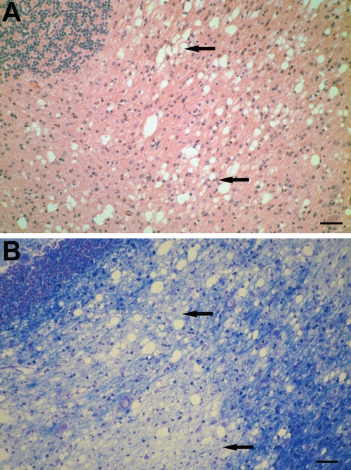
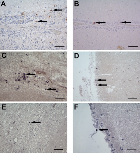
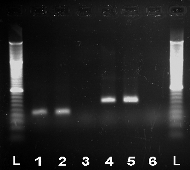
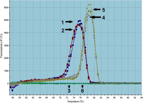
Leukoencephalopathies in dogs encompass presumably inherited conditions such as leukodystrophies, hypomyelination or spongiform degeneration, but other causes, such as virus infections and toxic or nutritional factors, might also play a contributory role. In this report, we provide evidence of parvovirus infection and replication in the brains of five 6-week-old Cretan hound puppies suffering from a puppy shaker syndrome and leukoencephalopathy. Although these puppies belonged to two different litters, they were closely related, tracing back two generations to the same sire. Histologically, a mild to moderate lymphohistiocytic meningitis, with focal lymphohistiocytic leukoencephalitis in two animals, and a mild to moderate vacuolation with myelin loss, mainly in the white matter of the cerebellum was detected. Vacuolation was also found in the corpus callosum, fimbria hippocampi, mesencephalon, capsula interna, basal ganglia, and hypothalamus. By immunohistology and in situ hybridization, either parvoviral antigen, DNA, mRNA, or replicative intermediate DNA were detected in the cerebellum, hippocampus, periventricular areas, corpus callosum, cerebral cortex, medulla oblongata, and spinal cord. Parvovirus antigen, DNA, and mRNA were present in cells of the outer granular layer of the cerebellum and in periventricular cells, most likely representing spongioblasts, glial cells, neurons, endothelial cells, occasional macrophages, and ependymal cells. Sequencing revealed canine parvovirus type 2 stretches. Thus, an association of parvovirus infection with the leukoencephalopathy seems likely, possibly facilitated by a genetic predisposition due to the mode of inbreeding in this particular dog breed.
Figures
Similar articles
-
Canine parvovirus types 2c and 2b circulating in North American dogs in 2006 and 2007.J Clin Microbiol. 2007 Dec;45(12):4044-7. doi: 10.1128/JCM.01300-07. Epub 2007 Oct 10. J Clin Microbiol. 2007. PMID: 17928423 Free PMC article.
-
Subacute massive necrotizing myocarditis by canine parvovirus type 2 infection with diffuse leukoencephalomalacia in a puppy.Vet Pathol. 1999 Jan;36(1):77-80. doi: 10.1354/vp.36-1-77. Vet Pathol. 1999. PMID: 9921761
-
Parvovirus Infection Is Associated With Myocarditis and Myocardial Fibrosis in Young Dogs.Vet Pathol. 2017 Nov;54(6):964-971. doi: 10.1177/0300985817725387. Epub 2017 Aug 16. Vet Pathol. 2017. PMID: 28812526 Free PMC article.
-
Parvovirus infection in domestic companion animals.Vet Clin North Am Small Anim Pract. 2008 Jul;38(4):837-50, viii-ix. doi: 10.1016/j.cvsm.2008.03.008. Vet Clin North Am Small Anim Pract. 2008. PMID: 18501282 Review.
-
Update on Canine Parvoviral Enteritis.Vet Clin North Am Small Anim Pract. 2020 Nov;50(6):1307-1325. doi: 10.1016/j.cvsm.2020.07.008. Epub 2020 Sep 2. Vet Clin North Am Small Anim Pract. 2020. PMID: 32891439 Free PMC article. Review.
Cited by
-
Quantitative DTI metrics in a canine model of Krabbe disease: comparisons versus age-matched controls across multiple ages.Neuroradiol J. 2018 Apr;31(2):168-176. doi: 10.1177/1971400917733431. Epub 2018 Jan 19. Neuroradiol J. 2018. PMID: 29350082 Free PMC article.
-
Congenital spongiform leukodystrophy in 2 female littermate German shepherd puppies.J Vet Intern Med. 2024 May-Jun;38(3):1730-1736. doi: 10.1111/jvim.17055. Epub 2024 Mar 27. J Vet Intern Med. 2024. PMID: 38544400 Free PMC article.
-
Automated segmentation of the canine corpus callosum for the measurement of diffusion tensor imaging.Neuroradiol J. 2016 Feb;29(1):4-12. doi: 10.1177/1971400915610924. Epub 2015 Nov 17. Neuroradiol J. 2016. PMID: 26577603 Free PMC article.
-
The first outbreak of feline panleukopenia virus infection in captive Pallas's cats in Xining Wildlife Park.Front Vet Sci. 2024 Aug 29;11:1418553. doi: 10.3389/fvets.2024.1418553. eCollection 2024. Front Vet Sci. 2024. PMID: 39268516 Free PMC article.
-
Pathogen Screening for Possible Causes of Meningitis/Encephalitis in Wild Carnivores From Saxony-Anhalt.Front Vet Sci. 2022 Apr 7;9:826355. doi: 10.3389/fvets.2022.826355. eCollection 2022. Front Vet Sci. 2022. PMID: 35464387 Free PMC article.
References
-
- Agungpriyono, D. R., K. Uchida, H. Tabaru, R. Yamaguchi, and S. Tateyama. 1999. Subacute massive necrotizing myocarditis by canine parvovirus type 2 infection with diffuse leukoencephalomalacia in a puppy. Vet. Pathol. 36:77-80. - PubMed
-
- Alvares-Buylla, A., B. Seri, and F. Doetsch. 2002. Identification of neural stem cells in the adult vertebrate brain. Brain Res. Bull. 57:751-758. - PubMed
-
- Barah, F., P. J. Vallely, M. L. Chiswick, G. M. Cleator, and J. R. Kerr. 2001. Association of human parvovirus B19 infection with acute meningoencephalitis. Lancet 358:729-730. - PubMed
-
- Barah, F., P. J. Vallely, G. M. Cleator, and J. R. Kerr. 2003. Neurological manifestations of human parvovirus B19 infection. Rev. Med. Virol. 13:185-199. - PubMed
MeSH terms
Substances
LinkOut - more resources
Full Text Sources
