. 2010 Jun 30;30(26):8759-68.
doi: 10.1523/JNEUROSCI.5229-09.2010.

Retinal degeneration and failure of photoreceptor outer segment formation in mice with targeted deletion of the Joubert syndrome gene, Ahi1

Affiliations

Retinal degeneration and failure of photoreceptor outer segment formation in mice with targeted deletion of the Joubert syndrome gene, Ahi1

Jennifer E Westfall et al. J Neurosci. .

Abstract

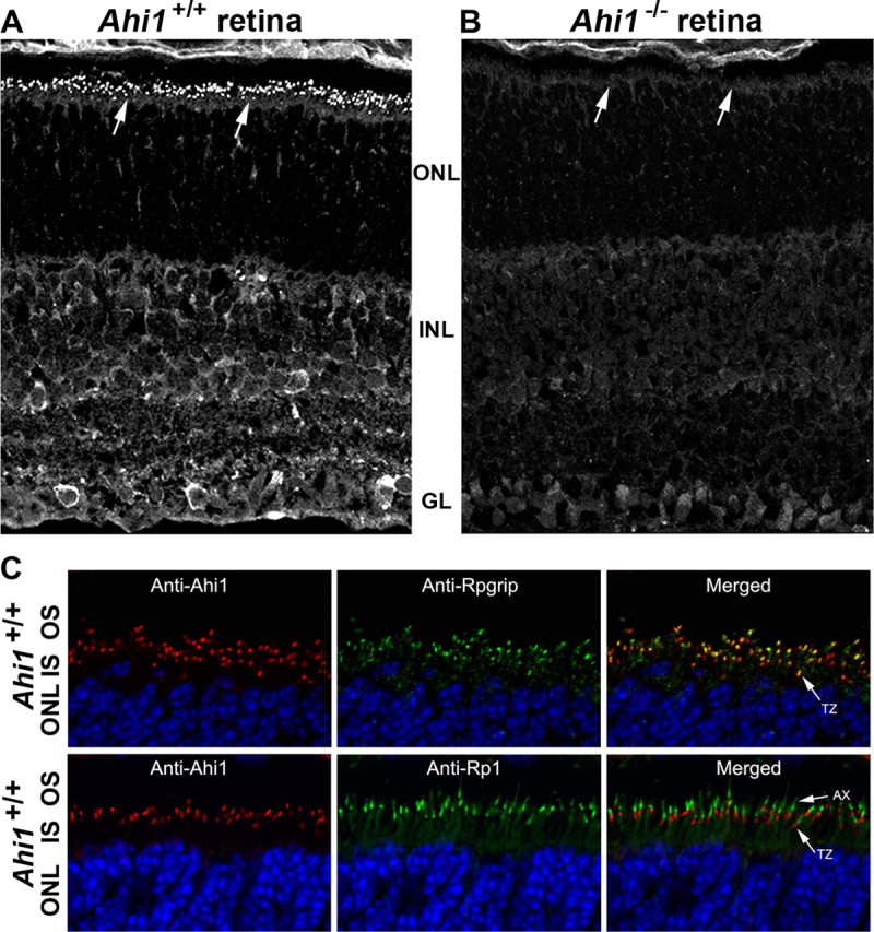
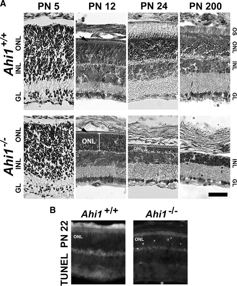
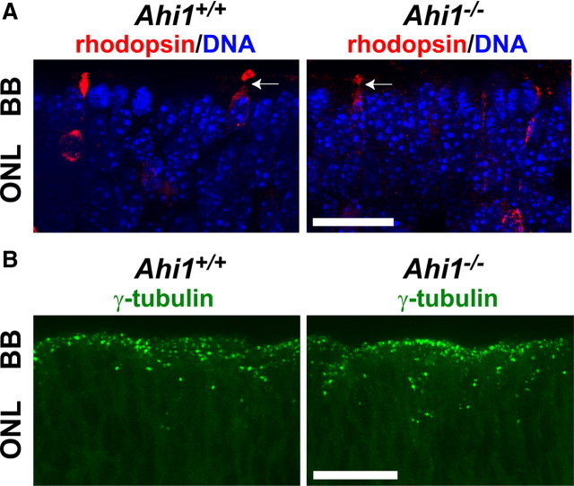
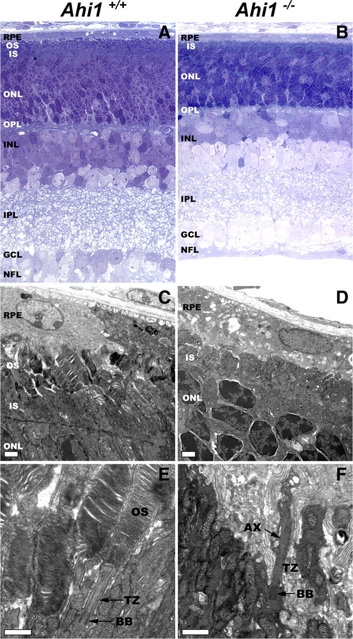
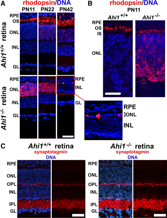
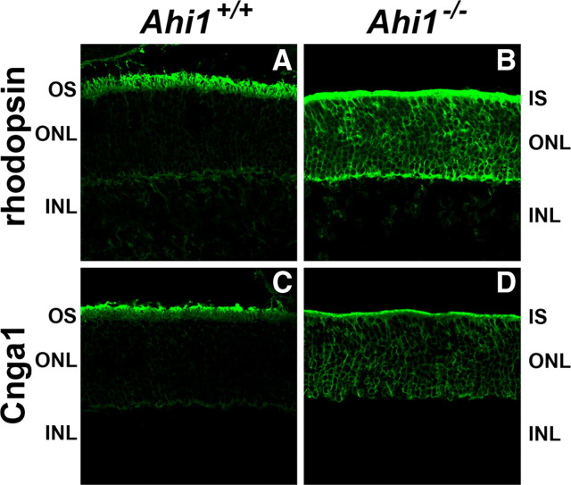
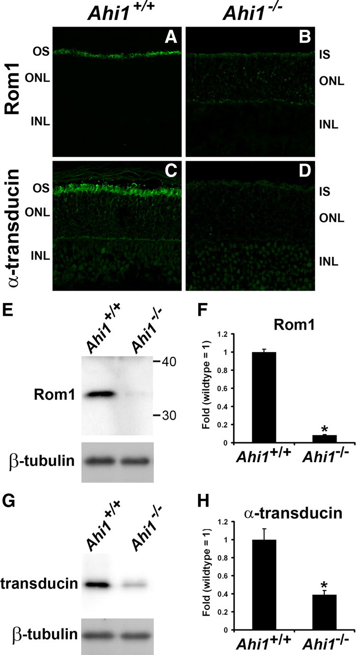
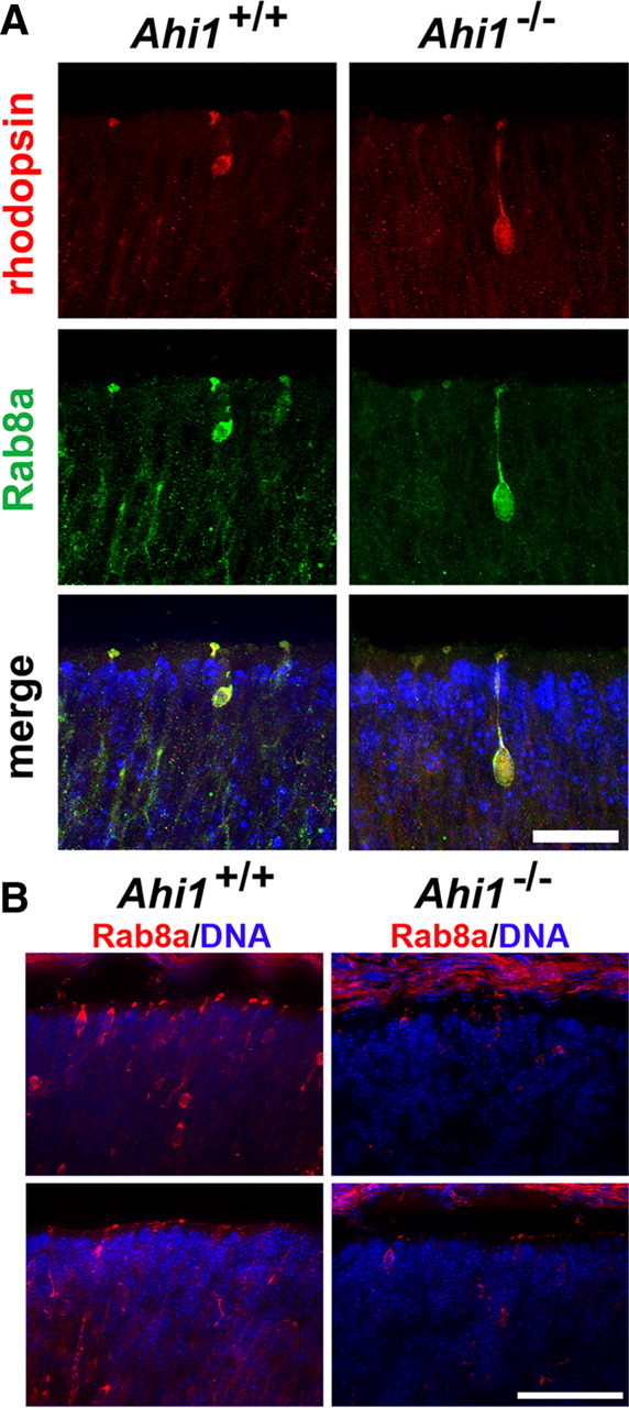
Vertebrate photoreceptors have a modified cilium composed of a basal body, axoneme and outer segment. The outer segment includes stacked membrane discs, containing opsin and the signal transduction apparatus mediating phototransduction. In photoreceptors, two distinct classes of vesicles are trafficked. Synaptic vesicles are transported down the axon to the synapse, whereas opsin-containing vesicles are transported to the outer segment. The continuous replacement of the outer segments imposes a significant biosynthetic and trafficking burden on the photoreceptors. Here, we show that Ahi1, a gene that when mutated results in the neurodevelopmental disorder, Joubert syndrome (JBTS), is required for photoreceptor sensory cilia formation and the development of photoreceptor outer segments. In mice with a targeted deletion of Ahi1, photoreceptors undergo early degeneration. Whereas synaptic proteins are correctly trafficked, photoreceptor outer segment proteins fail to be transported appropriately or are significantly reduced in their expression levels (i.e., transducin and Rom1) in Ahi1(-/-) mice. We show that vesicular targeting defects in Ahi1(-/-) mice are cilium specific, and our evidence suggests that the defects are caused by a decrease in expression of the small GTPase Rab8a, a protein required for accurate polarized vesicular trafficking. Thus, our results suggest that Ahi1 plays a role in stabilizing the outer segment proteins, transducin and Rom1, and that Ahi1 is an important component of Rab8a-mediated vesicular trafficking in photoreceptors. The retinal degeneration observed in Ahi1(-/-) mice recapitulates aspects of the retinal phenotype observed in patients with JBTS and suggests the importance of Ahi1 in photoreceptor function.


Figures

Figure 1.
Figure 1.
Ahi1 localization in the transition zone of the specialized cilium of the photoreceptors and in the inner retina. A, Immunostaining of Ahi1 in wild-type (Ahi1+/+) PN12 retinas. Arrows indicate targeting of Ahi1 to the ciliary transition zone. B, Lack of Ahi1 immunostaining in retina from age-matched mice that have a targeted deletion of Ahi1 (Ahi1−/−). Arrows indicate that lack of targeting of Ahi1 to the ciliary transition zone. C, PN12 wild-type (Ahi1+/+) mice with coimmunolabeling of Ahi1 (red) and the transition zone marker, Rpgrip (green in top row of images). Also labeled is the axoneme marker, Rp1 (green in bottom row of images). DNA is visualized with DAPI (blue). GL, Ganglion cell layer; INL, inner nuclear layer; ONL, outer nuclear layer; AX, axoneme; IS, inner segments; OS, outer segments; TZ, transition zone.
Figure 2.
Figure 2.
Retinal degeneration in the photoreceptors (outer nuclear layer) of mice with a targeted deletion of Ahi1. A, Cresyl violet staining of retinas from age-matched, wild-type mice (Ahi1+/+; top row) and mice with a targeted deletion of Ahi1 (Ahi1−/−; bottom row) at PN5, PN12, PN24, and PN200, demonstrating the progressive degeneration of the photoreceptor layer in Ahi1−/− retina, but not in Ahi1+/+ retina. The white brackets in the images from the Ahi1−/− retinas at PN12 and PN24 highlight the decrease in size of the photoreceptor layer. The black arrows are pointing to the outer segment layer in Ahi1+/+ retina and the absence of this layer in Ahi1−/− retina. Scale bar, 50 μm. B, TUNEL staining of apoptotic photoreceptor nuclei at PN22 in Ahi1−/− retinas (right), but not in Ahi1+/+ retinas (left). INL, Inner nuclear layer; IGL, ganglion cell layer; ONL, outer nuclear layer; OS, outer segments.
Figure 3.
Figure 3.
Basal bodies in retinal photoreceptors and early photoreceptor cell development were not abnormal in E18.5 Ahi1−/− mice. A, Rhodopsin-positive photoreceptor cells (red) in the outer retina extend processes that terminate in broadened segments in retinas from day E18.5 Ahi1+/+ and Ahi1−/− mice, characteristic of developing connecting cilia. Arrows are pointing to immature rhodopsin-positive outer membranes. DNA is visualized with DAPI (blue). B, Day E18.5 mouse retinas from Ahi1+/+ and Ahi1−/− mice were immunostained for the ciliary basal body (centriole) marker, γ-tubulin (green). Scale bars, 20 μm. BB, Basal bodies; ONL, outer nuclear layer.
Figure 4.
Figure 4.
Ahi1−/− mice fail to develop photoreceptor outer segments. A, B, Light micrographs of epoxy-embedded sections of central retinas from PN12 Ahi1+/+ (A) and Ahi1−/− (B) mice. The outer segments did not develop in retinas from Ahi1−/− mice. The thickness of the outer nuclear layer was reduced, compared with retinas from Ahi1+/+ littermate controls. C, D, Electron microscopic images showing the ultrastructure of photoreceptors from PN12 Ahi1+/+ (C) and Ahi1−/− (D) mice. Whereas the outer segments in the Ahi1+/+ retina had a normal appearance, the outer segments in the retinas from Ahi1−/− mice were completely absent. Scale bars, 2 μm. E, F, Details of the photoreceptor sensory cilium components, in retinas from PN12 Ahi1+/+ (E) and Ahi1−/− (F) mice. The stacks of nascent discs, the axoneme, basal body, and transition zone had a normal appearance in Ahi1+/+ mice. However, in the Ahi1−/− mice, although the axoneme, basal body, and transition zone appeared normal, no disc membranes were observed along the axoneme; instead, the axoneme was surrounded by disorganized membranous material at its distal end. Scale bars, 1 μm. AX, Axoneme; BB, basal body; INL, inner nuclear layer; IPL, inner plexiform layer; IS, inner segments; GCL, ganglion cell layer; NFL, nerve fiber layer; ONL, outer nuclear layer; OPL, outer plexiform layer; OS, outer segments; RPE, retinal pigment epithelium; TZ, transition zone.
Figure 5.
Figure 5.
Photoreceptor outer segments fail to mature in retinas from Ahi1−/− mice. A, Rhodopsin (red) is sparsely observed throughout the outer nuclear layer, but is highly present in photoreceptor outer segments in Ahi1+/+ mice (top row). The outer segments are shown elongating from PN11 to PN42. In the Ahi1−/− retina (bottom row), a thin line of rhodopsin immunostaining (red) can be seen along the photoreceptor inner segments; however, most of the rhodopsin has been mistargeted to the outer nuclear layer (ONL). Additionally, the ONL is thinner than in the wild type at PN22, and only a few rhodopsin-positive cells remain by PN42 (see the high-magnification image of the ONL in the Ahi1−/− retina). DNA is visualized with DAPI (blue). Scale bar, 50 μm. B, Higher magnification of rhodopsin immunostaining (red) in PN11 mouse retinas from Ahi1+/+ and Ahi1−/− mice. DNA is visualized with DAPI (blue). Scale bar, 20 μm. C, The synaptic vesicle marker synaptotagmin is not mistargeted in retinas from Ahi1−/− mice. PN11 retinas from Ahi1+/+ and Ahi1−/− mice were immunostained for synaptotagmin (red). Synaptotagmin staining is restricted primarily to the inner and outer plexiform layers in both Ahi1+/+ and Ahi1−/− mice. DNA is visualized with DAPI (blue). Scale bars, 50 μm. INL, Inner nuclear layer; IPL, inner plexiform layer; GL, ganglion cell layer; OPL, outer plexiform layer; OS, outer segments; RPE, retinal pigment epithelium.
Figure 6.
Figure 6.
Abnormal location of photoreceptor outer segment proteins in retinas from Ahi1−/− mice. A–D, Frozen sections of retinas from Ahi1+/+ and Ahi1−/− mice were labeled with antibodies to the photoreceptor outer segment proteins: rhodopsin (A, B) and Cnga1 (C, D). The outer segment proteins rhodopsin and Cnga1 were mistargeted to the inner segments and cell bodies of photoreceptor cells in Ahi1−/− retinas (B, D), compared to their outer segment location in the Ahi1+/+ control retinas (A, C). INL, Inner nuclear layer; IS, inner segments; ONL, outer nuclear layer; OS, outer segments.
Figure 7.
Figure 7.
Decreased levels of photoreceptor outer segment proteins in retinas from Ahi1−/− mice. A–D, Frozen sections of retinas from Ahi1+/+ and Ahi1−/− mice were labeled with antibodies to other photoreceptor outer segment proteins: Rom1 (A, B), and α-transducin (C, D). The outer segment protein Rom1 was mistargeted to the inner segments and cell bodies of photoreceptor cells in Ahi1−/− retinas (B) compared to their outer segment location in the Ahi1+/+ control retinas (A). Also, the levels of Rom1 and α-transducin were significantly reduced in Ahi1−/− retinas (A vs B, C vs D, respectively). E–H, These reductions in Rom1 and α-transducin levels in the photoreceptors were confirmed by Western blotting. The protein levels of endogenous Rom1 and α-transducin from dissected retinas from Ahi1+/+ and Ahi1−/− mice were analyzed by Western blotting (representative blots, E, G) and graphically displayed (F, H; n ≥ 3/genotype). The level of β-tubulin represents the loading control. The error bars represent SEM. Asterisks denote significance from Ahi1+/+ (Rom1, t6 = 23.55, p < 0.0001; α-transducin, t4 = 4.78, p < 0.001). INL, Inner nuclear layer; IS, inner segments; ONL, outer nuclear layer; OS, outer segments.
Figure 8.
Figure 8.
Reduced Rab8a expression in retinal photoreceptors from day E18.5 Ahi1−/− mice. A, Retinas from Ahi1+/+ and Ahi1−/− mice were immunostained for the membrane trafficking protein, Rab8a (green), and were colabeled with rhodopsin (red). Rab8a and rhodopsin colocalized in immature photoreceptor cells, in both Ahi1+/+ and Ahi1−/− mice. The same exposure settings were used for the Ahi1+/+ and Ahi1−/− retinas. DNA is visualized with DAPI (blue). Scale bar, 20 μm. B, Lower-magnification images of Rab8a (red) immunostaining in retinal photoreceptors from Ahi1+/+ and Ahi1−/− mice. Much weaker Rab8a immunostaining was observed in Ahi1−/− photoreceptors (right) than in Ahi1+/+ photoreceptors (left). The same exposure settings were used for the Ahi1+/+ and Ahi1−/− retinas. DNA is visualized with DAPI (blue). Scale bar, 50 μm.

Similar articles

Cited by

References

    1. Abd-El-Barr MM, Sykoudis K, Andrabi S, Eichers ER, Pennesi ME, Tan PL, Wilson JH, Katsanis N, Lupski JR, Wu SM. Impaired photoreceptor protein transport and synaptic transmission in a mouse model of Bardet-Biedl syndrome. Vision Res. 2007;47:3394–3407. - PMC - PubMed
    1. Adams NA, Awadein A, Toma HS. The retinal ciliopathies. Ophthalmic Genet. 2007;28:113–125. - PubMed
    1. Alfinito PD, Townes-Anderson E. Activation of mislocalized opsin kills rod cells: a novel mechanism for rod cell death in retinal disease. Proc Natl Acad Sci U S A. 2002;99:5655–5660. - PMC - PubMed
    1. Badano JL, Mitsuma N, Beales PL, Katsanis N. The ciliopathies: an emerging class of human genetic disorders. Annual review of genomics and human genetics. 2006;7:125–148. - PubMed
    1. Bascom RA, Manara S, Collins L, Molday RS, Kalnins VI, McInnes RR. Cloning of the cDNA for a novel photoreceptor membrane protein (rom-1) identifies a disk rim protein family implicated in human retinopathies. Neuron. 1992;8:1171–1184. - PubMed

Publication types

MeSH terms