. 2010 Dec;51(12):6441-7.
doi: 10.1167/iovs.10-5216. Epub 2010 Jun 30.

Multiplex cytokine analysis reveals elevated concentration of interleukin-8 in glaucomatous aqueous humor

Affiliations

Multiplex cytokine analysis reveals elevated concentration of interleukin-8 in glaucomatous aqueous humor

John Kuchtey et al. Invest Ophthalmol Vis Sci. 2010 Dec.

Abstract

Purpose: To test the hypothesis that immune activation occurs in glaucoma by comparing concentrations of multiple cytokines in aqueous humor (AH) from patients with primary open angle glaucoma (POAG) and from cataract patients without glaucoma as controls.

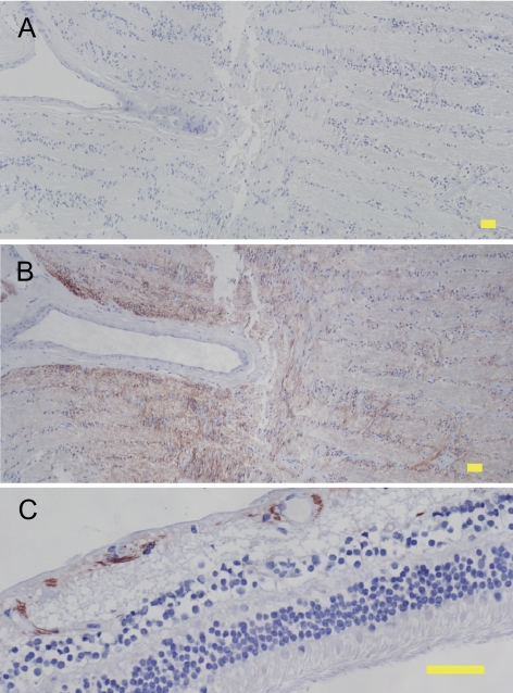
Methods: Cytokine concentrations in AH obtained during surgery were measured using microparticle-based immunoassays. Localized expression of IL-8 protein was investigated by immunohistochemistry of human eyes.

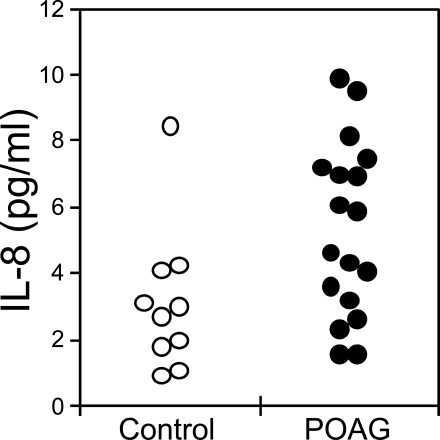
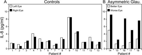
Results: Eight cytokines (IL-1β, IL-2, IL-4, IL-5, IL-10, IL-12, IFN-γ, and TNF-α) were below the limits of detection, and two cytokines (IL-18 and IL-15) were detected at low levels or in only a few patients. Although IL-6 was detected in 26 of 30 control patients (median, 2.7 pg/mL) and in 23 of 29 POAG patients (median, 1.6 pg/mL), the difference was not statistically significant. IL-8 was detected in 28 of 30 control patients (median, 1.8 pg/mL) and in all 29 POAG patients (median, 4.9 pg/mL). The higher IL-8 concentration in the AH of POAG patients was statistically significant (P < 0.001). In pairs of eyes from patients with asymmetric glaucomatous optic nerve damage, IL-8 concentration was higher in the AH of the more severely affected eye (P < 0.05). Patients with severe visual field defects had higher IL-8 concentrations in the AH than did patients with mild visual field defects. IL-8 protein expression was found in human retina and optic nerve.

Conclusions: Concentration of the inflammatory cytokine IL-8 is significantly elevated in the AH of POAG patients, supporting the hypothesis that immune activation occurs in glaucoma.


Figures

Figure 1.
Figure 1.
Concentrations of IL-6 (A) and IL-8 (B) in AH of patients without glaucoma (control, open circles, n = 30) and with POAG (POAG, closed circles, n = 29). Median concentrations for IL-6: control, 2.7 pg/mL; POAG, 1.6 pg/mL. Median concentrations for IL-8: control, 1.8 pg/mL; POAG, 4.9 pg/mL. IL-8 is significantly higher in POAG than in control patients (P < 0.001). Samples with cytokine concentration below the detection limit (0.5 pg/mL for IL-6 and 0.6 pg/mL for IL-8) are plotted on the bottom axis (0 pg/mL). Two control samples with IL-6 concentrations of 82 and 186 pg/mL are plotted on the top axis (>20 pg/mL).
Figure 2.
Figure 2.
Concentrations of IL-8 in AH of patients without glaucoma (control, open circles, n = 10) and with POAG (POAG, closed circles, n = 18) undergoing cataract surgery. Median concentrations: control, 2.9 pg/mL; POAG, 5.2 pg/mL. IL-8 is significantly higher in POAG than in control patients (P < 0.05).
Figure 3.
Figure 3.
IL-8 in AH from pairs of eyes of individual patients. IL-8 concentrations from the left eye versus the right eye are shown for individual control patients (A) and from the less severely affected eye (better eye) versus the more severely affected eye (worse eye) for individual glaucoma patients with clearly defined asymmetric glaucoma (B). Patients 14, 15, and 16 have POAG, and patients 17 and 18 have exfoliation glaucoma (B). The absolute value of the difference in IL-8 concentration (4.1 ± 2.23 pg/mL [mean ± SD]) for pairs of asymmetric glaucoma eyes was significantly greater than for control pairs (0.5 ± 0.36 pg/mL; P < 0.01).
Figure 4.
Figure 4.
IL-8 in AH of POAG patients with mild versus severe visual field defects. Concentrations of IL-8 in AH of patients with reliable HVF test results showing either mild (mild, open circles, n = 15) or severe (severe, closed circles, n = 15) visual field defects were compared. Median concentrations: mild, 4.7 pg/mL; severe, 7.3 pg/mL. IL-8 was significantly higher in patients with severe visual field defects than in patients with mild visual field defects (P < 0.05). Visual field defects were defined as mild for patients with mean deviations no worse than −6 dB and severe for mean deviations worse than −12 dB.
Figure 5.
Figure 5.
IL-8 protein expression in human cadaver eyes. Immunohistochemistry was performed on sections of paraformaldehyde-fixed human cadaver eyes using negative control IgG (A) or anti–IL-8 antibody (B, C) and detection with Nova Red substrate followed by hematoxylin counterstain. Reddish-brown: positive immunoreactivity. Blue: hematoxylin counterstain. Staining with negative control IgG resulted in no detectable immunoreactivity (A). IL-8 expression was found in the optic nerve (B) and inner retina (C). Yellow scale bar, 50 μm.

References

    1. Tezel G, Wax MB. The immune system and glaucoma. Curr Opin Ophthalmol. 2004;15:80–84 - PubMed
    1. Wax MB, Tezel G. Immunoregulation of retinal ganglion cell fate in glaucoma. Exp Eye Res. 2009;88:825–830 - PubMed
    1. Tezel G. The role of glia, mitochondria, and the immune system in glaucoma. Invest Ophthalmol Vis Sci. 2009;50:1001–1012 - PubMed
    1. Grus FH, Joachim SC, Wuenschig D, Rieck J, Pfeiffer N. Autoimmunity and glaucoma. J Glaucoma. 2008;17:79–84 - PubMed
    1. Yang J, Patil RV, Yu H, Gordon M, Wax MB. T cell subsets and sIL-2R/IL-2 levels in patients with glaucoma. Am J Ophthalmol. 2001;131:421–426 - PubMed

Publication types