. 2010 Oct 14;116(15):2803-11.
doi: 10.1182/blood-2010-03-276972. Epub 2010 Jun 30.

Live imaging of neutrophil motility in a zebrafish model of WHIM syndrome

Affiliations

Live imaging of neutrophil motility in a zebrafish model of WHIM syndrome

Kevin B Walters et al. Blood. .

Abstract

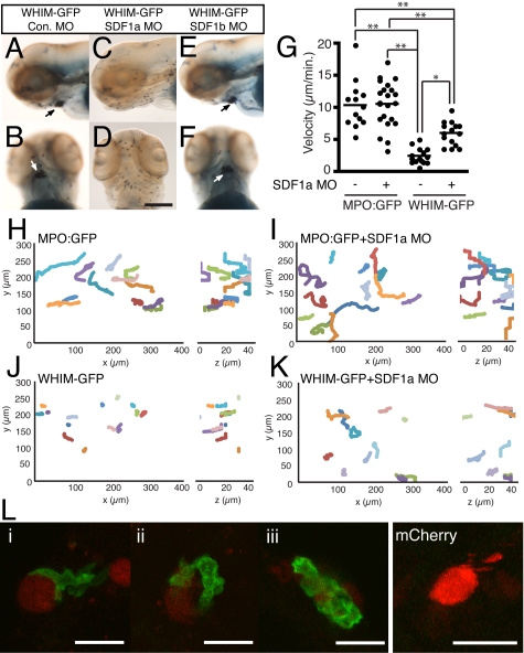
CXCR4 is a G protein-coupled chemokine receptor that has been implicated in the pathogenesis of primary immunodeficiency disorders and cancer. Autosomal dominant gain-of-function truncations of CXCR4 are associated with warts, hypo-gammaglobulinemia, infections, and myelokathexis (WHIM) syndrome, a primary immunodeficiency disorder characterized by neutropenia and recurrent infections. Recent progress has implicated CXCR4-SDF1 (stromal cell-derived factor 1) signaling in regulating neutrophil homeostasis, but the precise role of CXCR4-SDF1 interactions in regulating neutrophil motility in vivo is not known. Here, we use the optical transparency of zebrafish to visualize neutrophil trafficking in vivo in a zebrafish model of WHIM syndrome. We demonstrate that expression of WHIM mutations in zebrafish neutrophils induces neutrophil retention in hematopoietic tissue, impairing neutrophil motility and wound recruitment. The neutrophil retention signal induced by WHIM truncation mutations is SDF1 dependent, because depletion of SDF1 with the use of morpholino oligonucleotides restores neutrophil chemotaxis to wounds. Moreover, localized activation of a genetically encoded, photoactivatable Rac guanosine triphosphatase is sufficient to direct migration of neutrophils that express the WHIM mutation. The findings suggest that this transgenic zebrafish model of WHIM syndrome may provide a valuable tool to screen for agents that modify CXCR4-SDF1 retention signals.


Figures

Figure 1
Figure 1
Expression of CXCR4b and SDF1a in zebrafish larvae. (A) Reverse transcription–polymerase chain reaction of csf1r (macrophage marker), mpo (neutrophil marker), eflα (loading control), CXCR4a, and CXCR4b from MPO:Dendra2 high (hi) and low (lo) populations. WKM indicates whole kidney marrow from adult wild-type fish. (B-C) Whole-mount in situ hybridization of SDF1a expression in 2 dpf larvae, lateral view. Note SDF1a expression in the head (B) and CHT (C arrowheads). P indicates pronephric duct; cht, caudal hematopoietic tissue. (D) Whole-mount Sudan Black staining to visualize neutrophils in 2 dpf larvae, lateral view. Note neutrophil accumulation in areas of SDF1a expression in the head (box) and CHT (arrowheads). Bar = 200 μm (B-D).
Figure 2
Figure 2
Generation of WHIM-GFP transgenic larvae. (A) Alignment of the C-terminal tails of human CXCR4 with zebrafish CXCR4b and WHIM-truncated zebrafish CXCR4b. Note conservation of serine residues. Arrow marks an identified WHIM truncation mutation. (B) Fluorescence images of human embryonic kidney cells expressing GFP (first column), CXCR4b-GFP (second column), or WHIM-GFP (third column) after incubation with human SDF1 (bottom row) or vehicle control (top row). (C) Schematic of Tol2-MPO:zCXCR4b-WHIM-GFP vector injected to generate WHIM-GFP transgenic lines. (D) Schematic of 3-dpf zebrafish larvae. Boxed region is approximate area magnified in panel E. (E) Fluorescence image of the CHT region of a 3-dpf WHIM-GFP larvae. (F-G) High-magnification image of GFP-expressing neutrophils from the CHT of a MPO:GFP (F) and a WHIM-GFP (G, blow up of box in larva from panel E. Note membrane expression in panel G. Bar = 50 μm (E); 20 μm (B); 10 μm (F-G).
Figure 3
Figure 3
Neutrophil development in WHIM-GFP larvae. (A-F) Sudan Black staining in the CHT of wild-type (WT) (A,C,E) or WHIM-GFP (B,D,F) larvae at 3 (A,B), 7 (C,D), and 13 (E,F) dpf. Lateral view, anterior to the left. Arrows point to neutrophils over the gut (C) or in clumps in the CHT (B,D,F). Arrowheads point to neutrophils along the dorsal ridge (C) or midline (F). (G-L) Lateral view (G,I,K) or ventral view (H,J,L) of the head of WT (G-J) or WHIM-GFP (K,L) 3-dpf larvae stained with Sudan Black (G,H,K,L) or for SDF1a expression (I,J) by WISH. Arrows point to area of dark SDF1a expression (I,J) or areas of neutrophil accumulation (K,L) on the ventral side of the head under the jaw. (M-P) Sudan Black staining to show neutrophils in the kidney (arrows) in 7 (M-N) and 13 (O-P) dpf WT (M,O) or WHIM-GFP (N,P) larvae. Lateral view, g indicates gut. Bar = 200 μm (A-P).
Figure 4
Figure 4
Neutrophil retention in the head is SDF1a dependent. (A-F) Sudan Black–stained WHIM-GFP larvae injected with either control (A-B), SDF1a (C-D), or SDF1b (E-F) MO. Lateral (A,C,E) or ventral (B,D,F) view of the head at 3 dpf; arrows point to neutrophil accumulation. (G) The mean velocity of tracked neutrophils from the ventral head of uninjected or SDF1a morphant MPO:GFP or WHIM-GFP larvae. **P < .001, *P < .01. (H-K) Neutrophil migration in the ventral head of MPO:GFP (H), SDF1a morphant MPO:GFP (I), WHIM-GFP (J), or SDF1a morphant WHIM-GFP (K) larvae was tracked in 3 dimensions. The tracks are plotted in 3-dimensional space and viewed in the xy-plane (left) or zy-plane (right). Units are in micrometers on each axis. Only tracks of neutrophils that lasted for ≥ 14 minutes and only the first 14 minutes of longer tracks are included. Tracks were taken from supplemental Video 1 with additional tracks in SDF1a morphant larvae from additional videos not shown. (L) WHIM-GFP larvae at 2 or 3 dpf injected with Tol2-CMV:SDF1a-2A-mCherry at the 1-cell stage. Three examples of WHIM-GFP neutrophils (green) in close association with cells expressing SDF1a-2A-mcherry (red) in the body (i), head (ii), and yolk sac (iii). Cells expressing mCherry alone did not recruit WHIM-GFP neutrophils. Bar = 200 μm (A-F); 25 μm (L).
Figure 5
Figure 5
WHIM-GFP neutrophils fail to enter the blood stream and respond to wounding in the ventral tailfin. (A) Schematic of the tail of 3-dpf larvae and sample GFP frame from supplemental Video 3; red box is area of dorsal aorta where time-lapse imaging was performed. DA indicates dorsal aorta outlined by white lines, arrows indicate direction of blood flow; CHT, where GFP+ neutrophils not in circulation can be seen. White arrowheads indicate neutrophils in the circulation. (B) Quantification of neutrophils in the blood of MPO:GFP and WHIM-GFP 3-4 dpf transgenic larvae; *P < .01. Each dot represents a separate larva whose blood was analyzed by time-lapse imaging for 1 minute as in supplemental Video 3. (C) Time-lapse imaging of the wound response in WHIM-GFP transgenic larvae (from supplemental Video 4), GFP fluorescence overlaid with differential interference contrast image at indicated time points. (D-E) Sudan Black staining to show neutrophils at wounds in the ventral tailfin in 3-dpf control (D) or WHIM-GFP (E) 2 hours after wound. (F) Quantification of neutrophil recruitment to wounds in fixed larvae as in panels D and E; *P < .001; n = the number of individual larva wounded and counted; control = GFP siblings of WHIM-GFP larvae. (G) Time course of neutrophil wound recruitment in MPO:GFP and WHIM-GFP transgenic larvae. Error bars = SEM. **P < .001; *P < .01; n = 20-25 larvae at each time point. Bars = 200 μm (C-E); 20 μm (A).
Figure 6
Figure 6
WHIM-GFP neutrophils fail to respond to tail transections or chronic inflammatory signals. (A) Sudan Black staining of tails from Control (left) or WHIM-GFP (right) larvae injected with clint-ex1 MO to induce epidermal hyperproliferation and chronic inflammation in the tail. (B) Sudan Black staining of tail transections in Control (left) or WHIM-GFP (right) larvae. (C) Confocal imaging at wounds in WHIM-GFP (top) or Control (bottom) larvae at 3 dpf immunolabeled with a rabbit antibody to MPO and a fluorescein isothiocyanate–conjugated anti–rabbit Fab fragment (left) followed by a rhodamine-red–conjugated rabbit antibody to L-plastin (middle). Overlapping signals are yellow in the overlay (right). Arrows are MPO+, L-plastin+ neutrophils; arrowheads are MPO, L-plastin+ macrophages; white * indicates location of the wound. Control = GFP siblings of WHIM-GFP larvae. Representative images of ≥ 20 larvae in each condition. Bars = 200 μm (A-B); 100 μm (C).
Figure 7
Figure 7
Depletion of SDF1a and photoactivation of Rac are sufficient to restore WHIM-GFP neutrophil–directed migration in vivo. (A) Time-lapse imaging (from supplemental Video 10) of GFP fluorescence showing WHIM-GFP neutrophils responding to a wound (*) in the ventral tailfin in a 3-dpf SDF1a morphant larvae. White arrows indicate WHIM-GFP neutrophils. (B-C,E-F) Sudan Black staining of neutrophil response to wounding (* or line) in the ventral tailfin (B-C) or to tail transection (E-F) of 3-dpf WHIM-GFP transgenic larvae injected with control (B,E) or SDF1a (C,F) MO. (D) Quantification of WHIM-GFP neutrophil response in wounded 3-dpf morphant larvae fixed 2 hours after wound as in panels B and C; *P < .01. (G) Quantification of WHIM-GFP neutrophil response to tail transection in 3-dpf morphant larvae fixed 2 hours after transection as in panels E and F; *P < .05. (H-J) Laser stimulation with a 458-nm light induces directed migration of WHIM-GFP neutrophils also expressing mCherry-PA-Rac from the CHT. Repeated photoactivation was used to direct a single WHIM-GFP/mCherry-PA-Rac neutrophil away from the cell aggregate in the CHT into the tailfin. (H) Z-stack images from the indicated time points in supplemental Video 11 were summed into a single 2-dimensional image and then consolidated into a semi–1-dimensional line. Stars indicate time points and position of laser stimulations. (I) Two examples of the WHIM-GFP/mCherry-PA-Rac neutrophil protruding after stimulation from supplemental Video 11. Black circles indicate position of laser stimulation; white circles are included as reference points. (J) Composite differential interference contrast image of the posterior CHT, blood vessels, and tailfin from supplemental Videos 11 and 12 overlaid with the track (black line) of the directed WHIM-GFP/mCherry-PA-Rac neutrophil migration away from and return to the CHT. The starting and stopping points of photoactivation are indicated. Note that after termination of photoactivation the neutrophil immediately returns to the neutrophil aggregate in the CHT. Similar observations were made in 3 different experiments with 3 different larvae. CHT indicates caudal hematopoietic tissue; BV, blood vessel. Arrows indicate direction of migration. Bars = 200 μm (B-D,F); 100 μm (A); 40 μm (J); and 20 μm (I).

Comment in

  • A WHIM-sical zebrafish.
    Hickstein DD, West RR. Hickstein DD, et al. Blood. 2010 Oct 14;116(15):2621-2. doi: 10.1182/blood-2010-07-296426. Blood. 2010. PMID: 20947686 Free PMC article.

Similar articles

Cited by

References

    1. Bleul CC, Fuhlbrigge RC, Casasnovas JM, Aiuti A, Springer TA. A highly efficacious lymphocyte chemoattractant, stromal cell-derived factor 1 (SDF-1). J Exp Med. 1996;184(3):1101–1109. - PMC - PubMed
    1. Doitsidou M, Reichman-Fried M, Stebler J, et al. Guidance of primordial germ cell migration by the chemokine SDF-1. Cell. 2002;111(5):647–659. - PubMed
    1. Alkhatib G, Berger EA. HIV coreceptors: from discovery and designation to new paradigms and promise. Eur J Med Res. 2007;12(9):375–384. - PubMed
    1. Gelmini S, Mangoni M, Serio M, Romagnani P, Lazzeri E. The critical role of SDF-1/CXCR4 axis in cancer and cancer stem cells metastasis. J Endocrinol Invest. 2008;31(9):809–819. - PubMed
    1. Furze RC, Rankin SM. Neutrophil mobilization and clearance in the bone marrow. Immunology. 2008;125(3):281–288. - PMC - PubMed

Publication types

MeSH terms