

Spectrum of intracranial parenchymal lesions in patients with human immunodeficiency virus infection in the Republic of Korea
- PMID: 20592890
- PMCID: PMC2890875
- DOI: 10.3346/jkms.2010.25.7.1005
Spectrum of intracranial parenchymal lesions in patients with human immunodeficiency virus infection in the Republic of Korea
Abstract
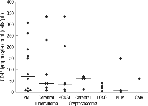
The incidence of specific intracranial parenchymal lesions of HIV-infected patients varies considerably between countries. In the Republic of Korea, the number of HIV-infected patients is increasing, but little is known regarding the spectrum of intracranial parenchymal lesions in these patients. The aim of the present study was to obtain this information. To identify HIV patients with intracranial parenchymal lesions, the electronic database of radiological reports for 1,167 HIV-infected patients, seen from 1999 to 2008 at the Seoul National University Hospital, were reviewed. Neuroradiologic studies were performed on 165 of these patients, and intracranial parenchymal lesions were detected in 40 (3.4%) of them. Thirty-seven were male, and median age was 41 yr (range, 26-61). At the time of the diagnosis of intracranial parenchymal lesions, median CD4(+) lymphocyte count was 40 cells/microL (range 5-560) and in 33 (82.5%) patients, it was less than 200 cells/microL. Progressive multifocal leukoencephalopathy (12 patients) is the most frequent intracranial parenchymal lesions, followed by intracranial tuberculoma (7 patients), primary central nervous system lymphoma (7 patients), intracranial cryptococcoma (4 patients), Toxoplasma encephalitis (4 patients), and disseminated non-tuberculous mycobacterial infection (3 patients).
Keywords: Acquired Immunodeficiency Syndrome; Brain Diseases; HIV; Korea.
Figures
References
-
- Levy RM, Bredesen DE, Rosenblum ML. Neurological manifestations of the acquired immunodeficiency syndrome (AIDS): experience at UCSF and review of the literature. J Neurosurg. 1985;62:475–495. - PubMed
-
- De Girolami U, Smith TW, Henin D, Hauw JJ. Neuropathology of the acquired immunodeficiency syndrome. Arch Pathol Lab Med. 1990;114:643–655. - PubMed
-
- Jellinger KA, Setinek U, Drlicek M, Bohm G, Steurer A, Lintner F. Neuropathology and general autopsy findings in AIDS during the last 15 years. Acta Neuropathol. 2000;100:213–220. - PubMed
-
- Masliah E, DeTeresa RM, Mallory ME, Hansen LA. Changes in pathological findings at autopsy in AIDS cases for the last 15 years. AIDS. 2000;14:69–74. - PubMed
-
- Davies J, Everall IP, Weich S, Glass J, Sharer LR, Cho ES, Bell JE, Majteny C, Gray F, Scaravilli F, Lantos PL. HIV-associated brain pathology: a comparative international study. Neuropathol Appl Neurobiol. 1998;24:118–124. - PubMed
MeSH terms
LinkOut - more resources
Full Text Sources
Medical
Research Materials
