

Successful resection of a giant left ventricular pseudoaneurysm developed later after mitral valve replacement
- PMID: 20592903
- PMCID: PMC2890888
- DOI: 10.3346/jkms.2010.25.7.1080
Successful resection of a giant left ventricular pseudoaneurysm developed later after mitral valve replacement
Abstract
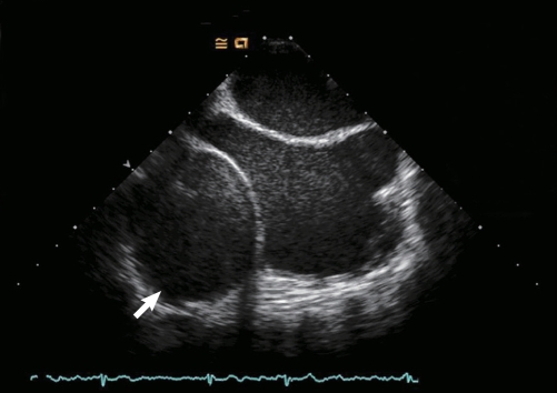
We present a case of successful surgical resection of a giant left ventricular (LV) pseudoaneurysm that developed 5 yr after mitral valve replacement (MVR). A 59-yr-old female was admitted with exertional chest pain radiating to left arm and back. 64-slice multidetector computed tomography (MDCT) revealed significant stenosis on the ostium of the first diagonal branch of the left anterior descending coronary artery and also a huge pseudoaneurysm compressing the right atrium and the inferior vena cava. She underwent resection of the pseudoaneurysm, and the pseudoaneurysm tunnel was repaired from the inside of LV cavity by removing the previously inserted prosthetic valve, followed by redo MVR together with coronary arterial bypass grafting (CABG) for a single-vessel disease. At the 6-month follow-up, the patient continued to do well without any complications.
Keywords: Aneurysm, False; Mitral Valve; Postoperative Complications.
Figures


References
-
- Miura T, Yamazaki K, Kihara S, Saito S, Miyagishima M, Aomi S, Kurosawa H. Transatrial repair of submitral left ventricular pseudoaneurysm. Ann Thorac Surg. 2008;85:643–645. - PubMed
-
- Schreurs M, Herregods MC, Bogaert J, Troost E, Budts W. Pseudoaneurysm of the left ventricle with moderate left-to-right shunt. Int J Cardiol. 2008;130:11–13. - PubMed
-
- Frances C, Romero A, Grady D. Left ventricular pseudoaneurysm. J Am Coll Cardiol. 1998;32:557–561. - PubMed
-
- Amasyali B, Cansel M, Kose S, Kursaklioglu H, Genc C, Isik E. Giant pseudoaneurysm of the left ventricle presenting 15 years after blunt chest trauma with high lateral myocardial infarction due to diagonal artery compression. Int J Cardiol. 2007;119 - PubMed
-
- Kollar A, Byrd BF, 3rd, Lui HK, Drinkwater DC., Jr Mitral valve replacement and endocavitary patch repair for a giant left ventricular pseudoaneurysm. Ann Thorac Surg. 2001;71:2020–2022. - PubMed
Publication types
MeSH terms
LinkOut - more resources
Full Text Sources
Medical
