

Prenatally detected congenital perineal mass using 3D ultrasound which was diagnosed as lipoblastoma combined with anorectal malformation: case report
- PMID: 20592907
- PMCID: PMC2890892
- DOI: 10.3346/jkms.2010.25.7.1093
Prenatally detected congenital perineal mass using 3D ultrasound which was diagnosed as lipoblastoma combined with anorectal malformation: case report
Abstract
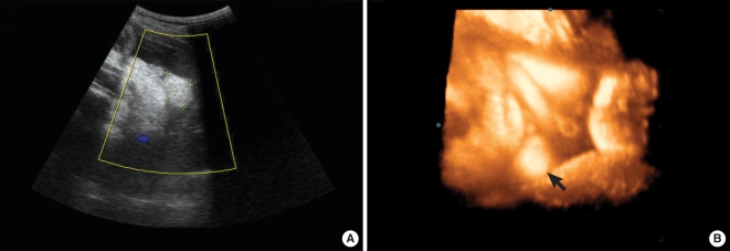
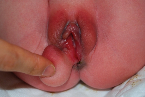
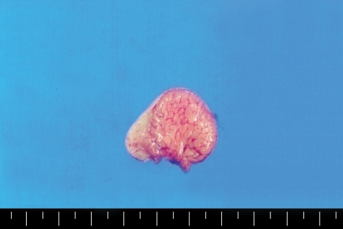
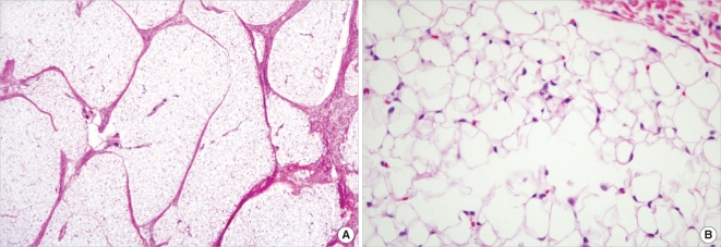
We report a case of prenatally diagnosed congenital perineal mass which was combined with anorectal malformation. The mass was successfully treated with posterior sagittal anorectoplasty postnatally. On ultrasound examination at a gestational age of 23 weeks the fetal perineal mass were found on the right side. Any other defects were not visible on ultrasonography during whole gestation. Amniocentesis was performed to evaluate the fetal karyotyping and acetylcholinesterase which were also normal. As the fetus grew up, the mass size was slowly increased more and more. At birth, a female neonate had a perineal mass on the right side as expected. During operation, the anal sphincteric displacement was found near the mass and reconstructed through posterior sagittal incision. This is the first reported case of prenatally diagnosed congenital perineal mass, after birth which was diagnosed as lipoblastoma and even combined with anorectal malformation. This case shows that it can be of clinical importance to be aware of this rare fetal perineal mass in prenatal diagnosis and counseling.
Keywords: Anal Canal; Congenital Abnormalities; Lipoblastoma; Perineum; Prenatal Diagnosis.
Figures
Similar articles
-
Infracoccygeal/transperineal window: new method to prenatally diagnose and classify level of anal atresia.Ultrasound Obstet Gynecol. 2024 Oct;64(4):521-527. doi: 10.1002/uog.29094. Epub 2024 Sep 1. Ultrasound Obstet Gynecol. 2024. PMID: 39219041
-
Prenatal diagnosis and neonatal management of anorectal malformation--a case report.Prague Med Rep. 2009;110(3):255-60. Prague Med Rep. 2009. PMID: 19655703
-
Fetal diagnosis of penoscrotal transposition associated with perineal lipoma in one twin.Fetal Diagn Ther. 2010;27(3):164-7. doi: 10.1159/000295807. Epub 2010 Mar 10. Fetal Diagn Ther. 2010. PMID: 20215739
-
[Perianal ultrasound].Z Gastroenterol. 2008 Jun;46(6):625-30. doi: 10.1055/s-2008-1027518. Z Gastroenterol. 2008. PMID: 18537090 Review. German.
-
First trimester expression of anorectal malformation: Case report and review of the literature.J Clin Ultrasound. 2018 Nov;46(9):591-597. doi: 10.1002/jcu.22612. Epub 2018 Sep 19. J Clin Ultrasound. 2018. PMID: 30229929 Review.
Cited by
-
Perineal lipoblastoma: a case report and review of literature.Int J Clin Exp Pathol. 2014 May 15;7(6):3370-4. eCollection 2014. Int J Clin Exp Pathol. 2014. PMID: 25031762 Free PMC article. Review.
-
Accessory scrotum with perineal lipoma diagnosed prenatally: case report and review of the literature.Nagoya J Med Sci. 2015 Aug;77(3):501-6. Nagoya J Med Sci. 2015. PMID: 26412897 Free PMC article.
-
Congenital perineal hamartomas with rectal duplication: A case report.Front Med (Lausanne). 2023 Jan 12;9:1066559. doi: 10.3389/fmed.2022.1066559. eCollection 2022. Front Med (Lausanne). 2023. PMID: 36714148 Free PMC article.
-
Infantile fibrosarcoma of the perineum with dorsal metastasis in a neonate: a case report original.BMC Pediatr. 2023 Jun 29;23(1):327. doi: 10.1186/s12887-023-04129-4. BMC Pediatr. 2023. PMID: 37386422 Free PMC article.
-
Anorectal Malformations Associated With Labioscrotal Fold Malformation and Perineal Mass in Pediatric Patients: Over a Decade of Experience.Front Pediatr. 2021 Feb 10;9:627188. doi: 10.3389/fped.2021.627188. eCollection 2021. Front Pediatr. 2021. PMID: 33643976 Free PMC article.
References
-
- Talwar MB, Misra K, Marya SK, Dev G. Fine needle aspiration cytology of a lipoblastoma. Acta Cytol. 1990;34:855–857. - PubMed
-
- Lopez-Ferrer P, Jimenez-Heffernan JA, Yebenes L, Vicandi B, Viguer JM. Fine-needle aspiration cytology of lipoblastoma: a report of two cases. Diagn Cytopathol. 2005;32:32–34. - PubMed
-
- Walaas L, Kindblom LG. Lipomatous tumors: a correlative cytologic and histologic study of 27 tumors examined by fine needle aspiration cytology. Hum Pathol. 1985;16:6–18. - PubMed
-
- Pollono DG, Tomarchio S, Drut R, Zaritzky M, Otero L, Vazquez AJ, Ripoll MC. Retroperitoneal and deep-seated lipoblastoma: diagnosis by ct scan and fine-needle aspiration biopsy. Diagn Cytopathol. 1999;20:295–297. - PubMed
-
- Jung SM, Chang PY, Luo CC, Huang CS, Lai JY, Hsueh C. Lipoblastoma/lipoblastomatosis: a clinicopathologic study of 16 cases in Taiwan. Pediatr Surg Int. 2005;21:809–812. - PubMed
Publication types
MeSH terms
LinkOut - more resources
Full Text Sources
Research Materials
