

The rhinolith-a possible differential diagnosis of a unilateral nasal obstruction
- PMID: 20592993
- PMCID: PMC2892702
- DOI: 10.1155/2010/845671
The rhinolith-a possible differential diagnosis of a unilateral nasal obstruction
Abstract
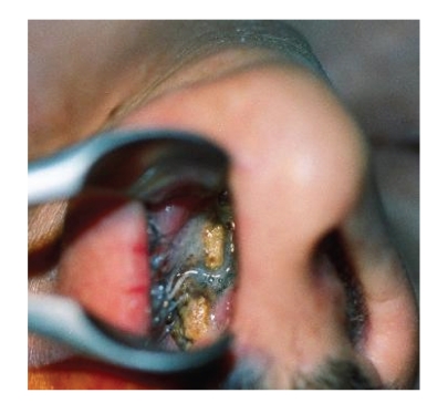
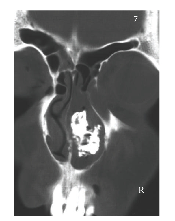
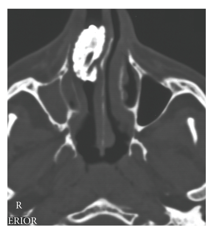
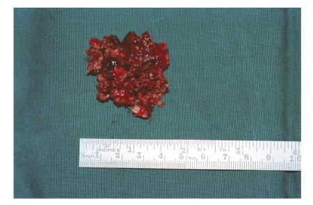
Rhinoliths are mineralised foreign bodies in the nasal cavity that are a chance finding at anterior rhinoscopy. Undiscovered, they grow appreciably in size and can cause a foul-smelling nasal discharge and breathing problems. Giant nasal stones are now a very rare occurrence, since improved diagnostic techniques, such as endoscopic/microscopic rhinoscopy, now make it possible to identify foreign bodies at an early stage of development. We report the case of a 37-year-old patient who, at the age of 5-6 years, introduced a foreign body, probably a stone, into his right nasal cavity. On presentation, he complained of difficulty in breathing through the right nostril that had persisted for the last 10 years. For the past four years a strong fetid smell from the nose had been apparent to those in his vicinity. Under general anaesthesia, the stone was removed in toto from the right nasal cavity. The possible genesis of the rhinolith is discussed, our case compared with those described in the literature, and possible differential diagnoses are considered.
Figures
References
-
- Denker A, Brünings W. Lehrbuch der Krankheiten des Ohres und der Luftwege. Jena, Germany: Gustav Fischer; 1912.
-
- Linnert D. Exogene Ursachen für die Entstehung von Nasensteinen. Zeitschrift fur Laryngologie, Rhinologie, Otologie und ihre Grenzgebiete. 1966;45(8):524–528. - PubMed
-
- Olbrich H. Rhinolith in einem Gaumendefekt. HNO. 1965;13:116–117. - PubMed
-
- Hoffmann AJ, Wagenfeld DJH. Rhinolith (nasal stone) associated with chronic otorrhea: a case report. South African Medical Journal. 1986;69(3):200–201. - PubMed
-
- Flood TR. Rhinolith: an unusual cause of palatal perforation. British Journal of Oral and Maxillofacial Surgery. 1988;26(6):486–490. - PubMed
Publication types
LinkOut - more resources
Full Text Sources
Miscellaneous
