

Defective NKT cell activation by CD1d+ TRAMP prostate tumor cells is corrected by interleukin-12 with α-galactosylceramide
- PMID: 20593019
- PMCID: PMC2892484
- DOI: 10.1371/journal.pone.0011311
Defective NKT cell activation by CD1d+ TRAMP prostate tumor cells is corrected by interleukin-12 with α-galactosylceramide
Abstract
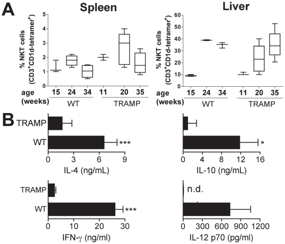
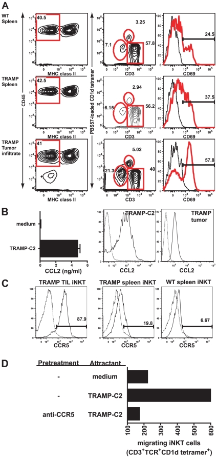
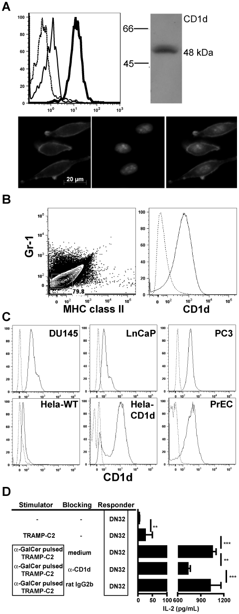
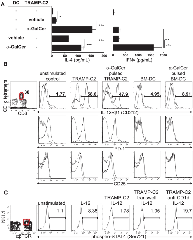
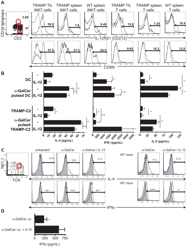
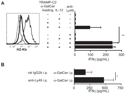
Numerical and functional defects of invariant natural killer T cells (iNKT) have been documented in human and mouse cancers, resulting in a defect in IFN production in several malignancies. iNKT cells recognize glycolipids presented on CD1d molecules by dendritic and related cells, leading to their activation and thereby regulating immune reactions. Activated iNKT cells cytokine secretion and cytotoxicity can inhibit existing and spontaneous tumor growth, progression, and metastasis. We have identified functional iNKT cell defects in the murine TRAMP prostate cancer model. We found that iNKT cells show the ability to migrate into TRAMP prostate tumors. This infiltration was mediated through CCL2: CCR5 chemokine: receptor interaction. Prostate tumor cells expressing CD1d partially activated iNKT cells, as appreciated by up-regulation of CD25, PD-1 and the IL-12R. However, despite inducing up-regulation of these activation markers and, hence, delivering positive signals, prostate tumor cells inhibited the IL-12-induced STAT4 phosphorylation in a cell-cell contact dependent but CD1d-independent manner. Consequently, tumor cells did not induce secretion of IFNgamma by iNKT cells. Blocking the inhibitory Ly49 receptor on iNKT cells in the presence of alpha-GalCer restored their IFNgamma production in vivo and in vitro. However, Ly49 blockade alone was not sufficient. Importantly, this defect could be also be reversed into vigorous secretion of IFNgamma by the addition of both IL-12 and the exogenous CD1d ligand alpha-galactosylceramide, but not by IL-12 alone, both in vivo and in vitro. These data underscore the potential to optimize iNKT-based therapeutic approaches.
Conflict of interest statement
Figures

Similar articles
-
Activation of human invariant natural killer T cells with a thioglycoside analogue of α-galactosylceramide.Clin Immunol. 2011 Aug;140(2):196-207. doi: 10.1016/j.clim.2011.03.016. Epub 2011 Apr 13. Clin Immunol. 2011. PMID: 21493160
-
The complementarity determining region 2 of BV8S2 (V beta 8.2) contributes to antigen recognition by rat invariant NKT cell TCR.J Immunol. 2006 Jun 15;176(12):7447-55. doi: 10.4049/jimmunol.176.12.7447. J Immunol. 2006. PMID: 16751390
-
Co-inhibitory roles for glucocorticoid-induced TNF receptor in CD1d-dependent natural killer T cells.Eur J Immunol. 2008 Aug;38(8):2229-40. doi: 10.1002/eji.200838167. Eur J Immunol. 2008. PMID: 18624295
-
Tailored design of NKT-stimulatory glycolipids for polarization of immune responses.J Biomed Sci. 2017 Mar 23;24(1):22. doi: 10.1186/s12929-017-0325-0. J Biomed Sci. 2017. PMID: 28335781 Free PMC article. Review.
-
Role of invariant natural killer T (iNKT) cells in systemic lupus erythematosus.Curr Med Chem. 2008;15(18):1778-87. doi: 10.2174/092986708785132988. Curr Med Chem. 2008. PMID: 18691038 Review.
Cited by
-
Targeting Natural Killer T Cells in Solid Malignancies.Cells. 2021 May 27;10(6):1329. doi: 10.3390/cells10061329. Cells. 2021. PMID: 34072042 Free PMC article. Review.
-
Natural Killer T Cells in Cancer Immunotherapy.Front Immunol. 2017 Sep 22;8:1178. doi: 10.3389/fimmu.2017.01178. eCollection 2017. Front Immunol. 2017. PMID: 29018445 Free PMC article. Review.
-
Optimizing iNKT-driven immune responses against cancer by modulating CD1d in tumor and antigen presenting cells.Clin Immunol. 2024 Dec;269:110402. doi: 10.1016/j.clim.2024.110402. Epub 2024 Nov 17. Clin Immunol. 2024. PMID: 39561929 Free PMC article. Review.
-
CAR-iNKT cells: redefining the frontiers of cellular immunotherapy.Front Immunol. 2025 Jul 11;16:1625426. doi: 10.3389/fimmu.2025.1625426. eCollection 2025. Front Immunol. 2025. PMID: 40718496 Free PMC article. Review.
-
The Dual Roles of Human γδ T Cells: Anti-Tumor or Tumor-Promoting.Front Immunol. 2021 Feb 16;11:619954. doi: 10.3389/fimmu.2020.619954. eCollection 2020. Front Immunol. 2021. PMID: 33664732 Free PMC article. Review.
References
-
- Bendelac A, Savage PB, Teyton L. The biology of NKT cells. Annu Rev Immunol. 2007;25:297–336. - PubMed
-
- Nowak M, Stein-Streilein J. Invariant NKT cells and tolerance. Int Rev Immunol. 2007;26:95–119. - PubMed
-
- Parekh VV, Wilson MT, Van Kaer L. iNKT-cell responses to glycolipids. Crit Rev Immunol. 2005;25:183–213. - PubMed
Publication types
MeSH terms
Substances
Grants and funding
LinkOut - more resources
Full Text Sources
Molecular Biology Databases
Miscellaneous
