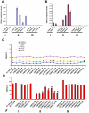
Variations of X chromosome inactivation occur in early passages of female human embryonic stem cells
- PMID: 20593031
- PMCID: PMC2892515
- DOI: 10.1371/journal.pone.0011330
Variations of X chromosome inactivation occur in early passages of female human embryonic stem cells
Abstract
X chromosome inactivation (XCI) is a dosage compensation mechanism essential for embryonic development and cell physiology. Human embryonic stem cells (hESCs) derived from inner cell mass (ICM) of blastocyst stage embryos have been used as a model system to understand XCI initiation and maintenance. Previous studies of undifferentiated female hESCs at intermediate passages have shown three possible states of XCI; 1) cells in a pre-XCI state, 2) cells that already exhibit XCI, or 3) cells that never undergo XCI even upon differentiation. In this study, XCI status was assayed in ten female hESC lines between passage 5 and 15 to determine whether XCI variations occur in early passages of hESCs. Our results show that three different states of XCI already exist in the early passages of hESC. In addition, we observe one cell line with skewed XCI and preferential expression of X-linked genes from the paternal allele, while another cell line exhibits random XCI. Skewed XCI in undifferentiated hESCs may be due to clonal selection in culture instead of non-random XCI in ICM cells. We also found that XIST promoter methylation is correlated with silencing of XIST transcripts in early passages of hESCs, even in the pre-XCI state. In conclusion, XCI variations already take place in early passages of hESCs, which may be a consequence of in vitro culture selection during the derivation process. Nevertheless, we cannot rule out the possibility that XCI variations in hESCs may reflect heterogeneous XCI states in ICM cells that stochastically give rise to hESCs.
Conflict of interest statement
Figures


Similar articles
-
Skewed X chromosome inactivation in diploid and triploid female human embryonic stem cells.Hum Reprod. 2009 Aug;24(8):1834-43. doi: 10.1093/humrep/dep126. Epub 2009 May 8. Hum Reprod. 2009. PMID: 19429659
-
X-inactivation in female human embryonic stem cells is in a nonrandom pattern and prone to epigenetic alterations.Proc Natl Acad Sci U S A. 2008 Mar 25;105(12):4709-14. doi: 10.1073/pnas.0712018105. Epub 2008 Mar 13. Proc Natl Acad Sci U S A. 2008. PMID: 18339804 Free PMC article.
-
The dynamic changes of X chromosome inactivation during early culture of human embryonic stem cells.Stem Cell Res. 2016 Jul;17(1):84-92. doi: 10.1016/j.scr.2016.05.011. Epub 2016 May 22. Stem Cell Res. 2016. PMID: 27262949
-
X chromosome inactivation in human pluripotent stem cells as a model for human development: back to the drawing board?Hum Reprod Update. 2017 Sep 1;23(5):520-532. doi: 10.1093/humupd/dmx015. Hum Reprod Update. 2017. PMID: 28582519 Review.
-
Regulation of X-chromosome inactivation by the X-inactivation centre.Nat Rev Genet. 2011 Jun;12(6):429-42. doi: 10.1038/nrg2987. Nat Rev Genet. 2011. PMID: 21587299 Review.
Cited by
-
Preventing erosion of X-chromosome inactivation in human embryonic stem cells.Nat Commun. 2022 May 6;13(1):2516. doi: 10.1038/s41467-022-30259-x. Nat Commun. 2022. PMID: 35523820 Free PMC article.
-
DNA methylation assay for X-chromosome inactivation in female human iPS cells.Stem Cell Rev Rep. 2011 Nov;7(4):969-75. doi: 10.1007/s12015-011-9238-6. Stem Cell Rev Rep. 2011. PMID: 21373884
-
Derivation of new human embryonic stem cell lines reveals rapid epigenetic progression in vitro that can be prevented by chemical modification of chromatin.Hum Mol Genet. 2012 Feb 15;21(4):751-64. doi: 10.1093/hmg/ddr506. Epub 2011 Nov 4. Hum Mol Genet. 2012. PMID: 22058289 Free PMC article.
-
Concise review: Pluripotency and the transcriptional inactivation of the female Mammalian X chromosome.Stem Cells. 2012 Jan;30(1):48-54. doi: 10.1002/stem.755. Stem Cells. 2012. PMID: 21997775 Free PMC article. Review.
-
Epigenetic reprogramming by naïve conditions establishes an irreversible state of partial X chromosome reactivation in female stem cells.Oncotarget. 2018 May 18;9(38):25136-25147. doi: 10.18632/oncotarget.25353. eCollection 2018 May 18. Oncotarget. 2018. PMID: 29861859 Free PMC article.
References
-
- Dvash T, Benvenisty N. Human embryonic stem cells as a model for early human development. Best Pract Res Clin Obstet Gynaecol. 2004;18:929–940. - PubMed
-
- Lu B, Malcuit C, Wang S, Girman S, Francis P, et al. Long-term Safety and Function of RPE from Human Embryonic Stem Cells in Preclinical Models of Macular Degeneration. Stem Cells 2009 - PubMed
-
- van Laake LW, Passier R, Monshouwer-Kloots J, Verkleij AJ, Lips DJ, et al. Human embryonic stem cell-derived cardiomyocytes survive and mature in the mouse heart and transiently improve function after myocardial infarction. Stem Cell Res. 2007;1:9–24. - PubMed
-
- Adewumi O, Aflatoonian B, Ahrlund-Richter L, Amit M, Andrews PW, et al. Characterization of human embryonic stem cell lines by the International Stem Cell Initiative. Nat Biotechnol. 2007;25:803–816. - PubMed
Publication types
MeSH terms
Substances
LinkOut - more resources
Full Text Sources
Molecular Biology Databases

