Review
. 2010;47(2):vii-xxxiv.
doi: 10.1682/jrrd.2010.02.0017.

Functional imaging and related techniques: an introduction for rehabilitation researchers

Review

Functional imaging and related techniques: an introduction for rehabilitation researchers

Bruce Crosson et al. J Rehabil Res Dev. 2010.

Abstract

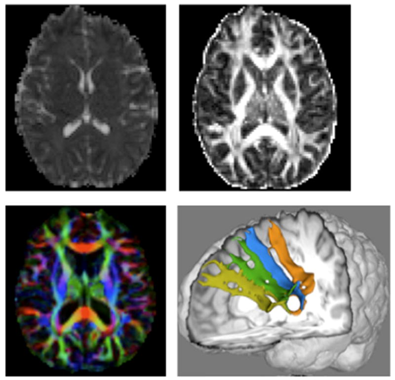
Functional neuroimaging and related neuroimaging techniques are becoming important tools for rehabilitation research. Functional neuroimaging techniques can be used to determine the effects of brain injury or disease on brain systems related to cognition and behavior and to determine how rehabilitation changes brain systems. These techniques include: functional magnetic resonance imaging (fMRI), positron emission tomography (PET), electroencephalography (EEG), magnetoencephalography (MEG), near infrared spectroscopy (NIRS), and transcranial magnetic stimulation (TMS). Related diffusion weighted magnetic resonance imaging techniques (DWI), including diffusion tensor imaging (DTI) and high angular resolution diffusion imaging (HARDI), can quantify white matter integrity. With the proliferation of these imaging techniques in rehabilitation research, it is critical that rehabilitation researchers, as well as consumers of rehabilitation research, become familiar with neuroimaging techniques, what they can offer, and their strengths and weaknesses The purpose to this review is to provide such an introduction to these neuroimaging techniques.


Figures

Figure 1
Figure 1
Hemodynamic response expressed as a percent change of the total signal. Note the positive phase of the response takes around 12 sec to resolve.
Figure 2
Figure 2
Pre- and post-treatment images for one subject who received the intention treatment. Red represents R2 ≥ .20; orange represents R2 ≥ .25. Note the lack of left frontal activity on post-compared to pre-treatment images, and the general reduction in frontal activity from pre to post treatment. Lateral frontal laterality indices are displayed at the bottom of the image, with 1.0 representing activity entirely lateralized to the left and -1.0 representing activity entirely lateralized to the right.
Figure 3
Figure 3
Schematic diagram for ASL imaging. The labeling of arterial blood is proximal to the tissue of interest, as shown by the blue plane at left. After a delay to let labeled blood to arrive at the tissue sites, imaging acquisitions will be performed. In some ASL techniques, control experiments will be done using the symmetric labeling RF pulse at the distal site to minimize the MT effects (light blue plane at right).
Figure 4
Figure 4
CBF maps (left) and asymmetry analysis results (right) from ASL perfusion study of a 76 year-old female stroke patient. Asymmetry index (%) = (CBF(right) - CBF(left))/(CBF(right)+CBF(left)) × 100. Specific perfusion territories are designated by ACA (anterior cerebral arteries), MCA (middle cerebral arteries), and PCA (posterior cerebral arteries). In the bar graph, the negative index value for ACA is indicated with a different pattern. In the CBF maps, brighter colors indicate greater CBF. Note the decreased CBF in the posterior right side of the images, consistent with the greater asymmetry in CBF for the posterior cerebral artery (PCA) distribution compared to the anterior cerebral artery (ACA) and middle cerebral artery (MCA).
Figure 5
Figure 5
DW-MRI measures from left to right, top row: mean diffusivity (MD) map, fractional anisotropy (FA) map; bottom row: FA map overlaid with the principle diffusion direction (PDD) in color (red left to right, blue inferior to superior, green anterior to posterior). Bottom right: 3-dimensional rendering of previously undocumented fiber paths connecting Broca’s area (Brodmann’s areas 44/45) with Brodmann’s area 9 (light green).
Figure 6
Figure 6
An example 1H spectrum, acquired at 3T, of the left basal ganglia of a normal control subject, post-processed in LCModel. Metabolites included in the modeled basis spectra are shown in the table on the right. Those detected with a reasonable confidence are in blue font. The %SD indicate Cramer-Rao lower error bounds.
Figure 7
Figure 7
Cortical motor map of first dorsal interosseous (FDI) muscle using single pulse TMS overlaid onto 3-Dimensional rendering of fMRI activation during FDI exercise. Site 74 (blue circle) represents cortical site of maximal sensitivity to FDI activation (aka the “hotspot”). Green circles are areas that also generate MEP in the target muscle, while red circles are the map’s boundary. Orange areas represents active areas during fMRI at F > 5.0 (p<.0001, uncorrected).

References

    1. Abutalebi J, Rosa PA, Tettamanti M, Green DW, Cappa SF. Bilingual aphasia and language control: a follow-up fMRI and intrinsic connectivity study. Brain Lang. 2009;109:141–156. - PubMed
    1. Babiloni C, Pizzella V, Gratta CD, Ferretti A, Romani GL. Fundamentals of electroencefalography, magnetoencefalography, and functional magnetic resonance imaging. Int Rev Neurobiol. 2009;86:67–80. - PubMed
    1. Barker PB, Soher BJ, Blackband SJ, Chatham JC, Mathews VP, Bryan RN. Quantitation of proton NMR spectra of the human brain using tissue water as an internal concentration reference. NMR Biomed. 1993;6:89–94. - PubMed
    1. Basser PJ, Mattiello J, LeBihan D. MR diffusion tensor spectroscopy and imaging. Biophys J. 1994;66:259–267. - PMC - PubMed
    1. Bengtsson SL, Nagy Z, Skare S, Forsman L, Forssberg H, Ullen F. Extensive piano practicing has regionally specific effects on white matter development. Nature Neuroscience. 2005;8:1148–1150. - PubMed

LinkOut - more resources