

Role of diffusion-weighted magnetic resonance imaging in the diagnosis of extrahepatic cholangiocarcinoma
- PMID: 20593506
- PMCID: PMC2896758
- DOI: 10.3748/wjg.v16.i25.3196
Role of diffusion-weighted magnetic resonance imaging in the diagnosis of extrahepatic cholangiocarcinoma
Abstract
Aim: To determine the clinical value of diffusion-weighted imaging (DWI) for the diagnosis of extrahepatic cholangiocarcinoma (EHCC) by comparing the diagnostic sensitivity of DWI and magnetic resonance cholangiopancreatography (MRCP).
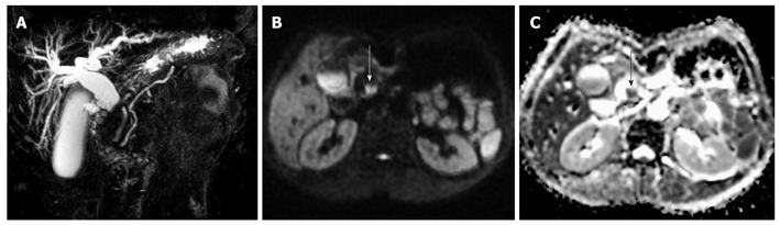
Methods: Magnetic resonance imaging examination was performed in 56 patients with suspected EHCC. T1-weighted imaging, T2-weighted imaging, MRCP and DWI sequence, DWI using single-shot spin-echo echo-planar imaging sequence with different b values (100, 300, 500, 800 and 1000 s/mm(2)), were performed. All cases were further confirmed by surgery or histopathological diagnosis. Two radiologists jointly performed the analysis of the DWI and MRCP images. Apparent diffusion coefficient (ADC) value and signal-noise ratio were calculated for EHCC. Sensitivity, specificity, accuracy, positive predictive value and negative predictive value were tested using DWI with a b value of 500 s/mm(2) and MRCP images, respectively.
Results: Histopathological diagnosis confirmed that among the 56 cases, 35 were EHCC (20 hilar and 15 distal extrahepatic), 16 were cholangitis, and 5 were calculus of bile duct. Thirty-three out of the 35 EHCC cases were detected by DWI. EHCC exhibited differential levels of high signal intensity in DWI and low signal intensity in the ADC map. The mean value for ADC was (1.31 +/- 0.29) x 10(-3) mm(2)/s. The detection rate of EHCC was significantly higher by DWI (94.3%) than by MRCP (74.3%) (P < 0.05). There was a significant difference in sensitivity (94.3% vs 74.3%), specificity (100% vs 71.4%), accuracy (96.4% vs 73.2%), positive predictive value (100% vs 81.3%), and negative predictive value (91.3% vs 62.5%) between DWI and MRCP in diagnosing EHCC.
Conclusion: DWI has a high sensitivity for the detection of EHCC as it shows the EHCC lesion more unambiguously than MRCP does. DWI can also provide additional clinically important information in EHCC patients when added to routine bile duct MR imaging protocols.
Figures
Similar articles
-
Diffusion-weighted MR imaging for detection of extrahepatic cholangiocarcinoma.Eur J Radiol. 2012 Nov;81(11):2961-5. doi: 10.1016/j.ejrad.2011.12.040. Epub 2012 Jan 28. Eur J Radiol. 2012. PMID: 22285604
-
Added value of diffusion-weighted imaging to MR cholangiopancreatography for the diagnosis of bile duct dilatations.Abdom Radiol (NY). 2016 Mar;41(3):485-92. doi: 10.1007/s00261-015-0612-8. Abdom Radiol (NY). 2016. PMID: 27039319
-
Value of diffusion-weighted MR imaging in the diagnosis of lymph node metastases in patients with cholangiocarcinoma.Abdom Radiol (NY). 2016 Oct;41(10):1937-41. doi: 10.1007/s00261-016-0791-y. Abdom Radiol (NY). 2016. PMID: 27271285
-
Hilar cholangiocarcinoma: MRI/MRCP in staging and treatment planning.Abdom Imaging. 2008 Jul-Aug;33(4):444-51. doi: 10.1007/s00261-007-9281-6. Abdom Imaging. 2008. PMID: 17638040 Review.
-
MRCP and 3D LAVA imaging of extrahepatic cholangiocarcinoma at 3 T MRI.Clin Radiol. 2012 Jun;67(6):579-86. doi: 10.1016/j.crad.2011.10.016. Epub 2011 Dec 3. Clin Radiol. 2012. PMID: 22137873 Review.
Cited by
-
Abdominal applications of diffusion-weighted magnetic resonance imaging: Where do we stand.World J Radiol. 2013 Mar 28;5(3):68-80. doi: 10.4329/wjr.v5.i3.68. World J Radiol. 2013. PMID: 23671743 Free PMC article.
-
Diffusion-Weighted Imaging with Two Different b-Values in Detection of Solid Focal Liver Lesions.Biomed Res Int. 2016;2016:8128207. doi: 10.1155/2016/8128207. Epub 2016 Feb 25. Biomed Res Int. 2016. PMID: 27019851 Free PMC article. Clinical Trial.
-
Biliary strictures: diagnostic considerations and approach.Gastroenterol Rep (Oxf). 2015 Feb;3(1):22-31. doi: 10.1093/gastro/gou072. Epub 2014 Oct 28. Gastroenterol Rep (Oxf). 2015. PMID: 25355800 Free PMC article. Review.
-
Evaluating Biliary Malignancy with Measured and Calculated Ultra-high b-value Diffusion-weighted MR Imaging at 3T.Magn Reson Med Sci. 2024 Oct 1;23(4):428-437. doi: 10.2463/mrms.mp.2022-0144. Epub 2023 May 13. Magn Reson Med Sci. 2024. PMID: 37183027 Free PMC article.
-
Imaging of Cholangiocarcinoma.Visc Med. 2016 Dec;32(6):402-410. doi: 10.1159/000453009. Epub 2016 Dec 6. Visc Med. 2016. PMID: 28229074 Free PMC article. Review.
References
-
- Bammer R, Schoenberg SO. Current concepts and advances in clinical parallel magnetic resonance imaging. Top Magn Reson Imaging. 2004;15:129–158. - PubMed
-
- Le Bihan D, Turner R, Douek P, Patronas N. Diffusion MR imaging: clinical applications. AJR Am J Roentgenol. 1992;159:591–599. - PubMed
-
- Koh DM, Collins DJ. Diffusion-weighted MRI in the body: applications and challenges in oncology. AJR Am J Roentgenol. 2007;188:1622–1635. - PubMed
-
- Stroszczynski C, Hunerbein M. Malignant biliary obstruction: value of imaging findings. Abdom Imaging. 2005;30:314–323. - PubMed
Publication types
MeSH terms
LinkOut - more resources
Full Text Sources
Medical
