

Surgical treatment of hepatocellular carcinoma with severe intratumoral arterioportal shunt
- PMID: 20593509
- PMCID: PMC2896761
- DOI: 10.3748/wjg.v16.i25.3211
Surgical treatment of hepatocellular carcinoma with severe intratumoral arterioportal shunt
Abstract
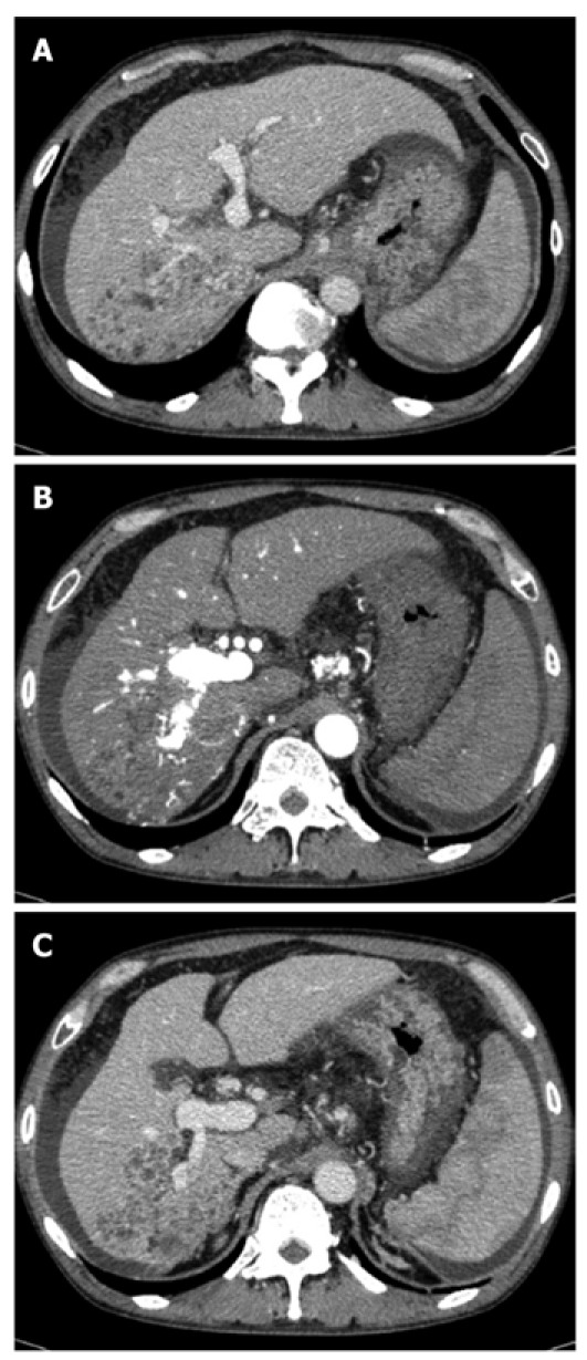
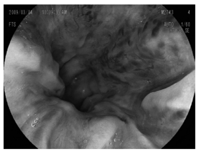
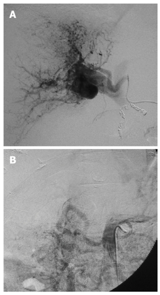
We report a case of hepatocellular carcinoma (HCC) that caused a severe arterioportal shunt (APS). A 49-year-old man was admitted to hospital due to esophagogastric variceal hemorrhage and HCC, and underwent endoscopic variceal ligation (EVL) and endoscopic injection sclerotherapy (EIS). He was then referred to our hospital. Abdominal computed tomography revealed a low-density lesion in the posterior segment of the liver and an intratumoral APS, which caused portal hypertension. Although the patient underwent EVL, EIS, Hassab's operation, and transcatheter arterial embolization for APS, he vomited blood due to rupture of esophagogastric varices. Right hepatectomy was performed for the treatment of HCC and APS, although the indocyanine green retention value at 15 min after intravenous injection was poor (30%). The patient's postoperative course was uneventful. Eventually, APS disappeared and the esophagogastric varices improved.
Figures
Similar articles
-
Additional Hassab's operation for esophagogastric varices in cirrhotic patients with resectable hepatocellular carcinoma.Hepatogastroenterology. 2008 Sep-Oct;55(86-87):1686-90. Hepatogastroenterology. 2008. PMID: 19102370
-
Treatment of concomitant gastric varices in patients with hepatocellular carcinoma at a single Japanese institute.Hepatogastroenterology. 2009 May-Jun;56(91-92):857-60. Hepatogastroenterology. 2009. PMID: 19621717
-
Major arterioportal shunt caused by hepatocellular carcinoma.J Nippon Med Sch. 2007 Aug;74(4):314-8. doi: 10.1272/jnms.74.314. J Nippon Med Sch. 2007. PMID: 17878703
-
Endoscopic variceal ligation compared with endoscopic injection sclerotherapy for treatment of esophageal variceal hemorrhage: a meta-analysis.World J Gastroenterol. 2015 Feb 28;21(8):2534-41. doi: 10.3748/wjg.v21.i8.2534. World J Gastroenterol. 2015. PMID: 25741164 Free PMC article. Review.
-
Endoscopic band ligation of oesophageal varices.Br J Surg. 1999 Apr;86(4):437-46. doi: 10.1046/j.1365-2168.1999.01109.x. Br J Surg. 1999. PMID: 10215811 Review.
Cited by
-
Surgical approach to multifocal hepatocellular carcinoma with portal vein thrombosis and arterioportal shunt leading to portal hypertension and bleeding: a case report.World J Surg Oncol. 2012 Feb 13;10:34. doi: 10.1186/1477-7819-10-34. World J Surg Oncol. 2012. PMID: 22330617 Free PMC article.
-
Short-Term Oral Sorafenib for Therapy of Intratumoral Shunts of Hepatocellular Carcinoma to Enable Intraarterial Treatment.Cardiovasc Intervent Radiol. 2019 Oct;42(10):1494-1499. doi: 10.1007/s00270-019-02294-7. Epub 2019 Jul 30. Cardiovasc Intervent Radiol. 2019. PMID: 31363899 Free PMC article.
References
-
- Lazaridis KN, Kamath PS. Images in hepatology. Arterio-portal fistula causing recurrent variceal bleeding. J Hepatol. 1998;29:142. - PubMed
-
- Morse SS, Sniderman KW, Galloway S, Rapoport S, Ross GR, Glickman MG. Hepatoma, arterioportal shunting, and hyperkinetic portal hypertension: therapeutic embolization. Radiology. 1985;155:77–82. - PubMed
-
- Velázquez RF, Rodríguez M, Navascués CA, Linares A, Pérez R, Sotorríos NG, Martínez I, Rodrigo L. Prospective analysis of risk factors for hepatocellular carcinoma in patients with liver cirrhosis. Hepatology. 2003;37:520–527. - PubMed
-
- Matsuyama K, Fukuda Y, Miyake H, Yogita S, Tashiro S. Experimental study of the evaluation of liver function on the opposite side during portacaval anastomosis and ligation of the left portal branch. J Med Invest. 2004;51:84–95. - PubMed
-
- Liver Cancer Study Group of Japan. General rules for the clinical and pathological study of primary liver cancer. 2nd English ed. Tokyo: Kanehara; 2003. pp. 13–14.
Publication types
MeSH terms
LinkOut - more resources
Full Text Sources
Medical
Miscellaneous
