Cervicofacial venous malformations. MRI features and interventional strategies
- PMID: 20594480
- PMCID: PMC3572475
- DOI: 10.1177/159101990200800302
Cervicofacial venous malformations. MRI features and interventional strategies
Abstract
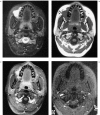
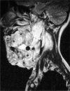
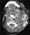
We retrospectively evaluated 53 consecutive patients with cervicofacial venous malformation who had sclerotherapy. This review included a demographic analysis, MRI reexamination and tabulation of interventional therapeutic strategies. All patients whose MRI studies were included in this review demonstrated characteristic findings: space occupying lesion with hyperintense T2 signal abnormality, patchy contrast enhancement, and no flow signal on the gradient echo images.We concluded that a complete MRI work-up of these patients requires post-contrast scanning and gradient-echo imaging in addition to the standard T1 and T2 weighted spin echo imaging. The majority of patients had sporadic (non-familial) venous anomalies. Sinus pericranii (SP) was identified in six patients (11%) and blue rubber bleb nevus syndrome (BRBNS) was found in two patients (4%). MRI findings of sinus pericranii are discussed in detail. Although sodium tetradecyl and/or absolute ethanol are the most commonly used sclerosants, a wide variety of therapeutic strategies (depending on the nature of the abnormality) are also needed for these patients.
Figures


Similar articles
-
Developmental venous anomalies and sinus pericranii in the blue rubber-bleb nevus syndrome. Case report.J Neurosurg. 2003 Aug;99(2):409-11. doi: 10.3171/jns.2003.99.2.0409. J Neurosurg. 2003. PMID: 12924718 Review.
-
Blue rubber bleb nevus syndrome in a Malay girl: A case report and literature review.Int J Surg Case Rep. 2020;72:202-206. doi: 10.1016/j.ijscr.2020.05.036. Epub 2020 Jun 1. Int J Surg Case Rep. 2020. PMID: 32544829 Free PMC article.
-
Pyogenic hepatic abscesses: MRI findings on T1- and T2-weighted and serial gadolinium-enhanced gradient-echo images.J Magn Reson Imaging. 1999 Feb;9(2):285-90. doi: 10.1002/(sici)1522-2586(199902)9:2<285::aid-jmri20>3.0.co;2-s. J Magn Reson Imaging. 1999. PMID: 10077026
-
Stomach diseases: MR evaluation using combined t2-weighted single-shot echo train spin-echo and gadolinium-enhanced spoiled gradient-echo sequences.J Magn Reson Imaging. 1999 Dec;10(6):950-60. doi: 10.1002/(sici)1522-2586(199912)10:6<950::aid-jmri7>3.0.co;2-h. J Magn Reson Imaging. 1999. PMID: 10581508
-
[Diagnostic imaging of hemangiomas in the brain].Brain Nerve. 2011 Jan;63(1):5-15. Brain Nerve. 2011. PMID: 21228443 Review. Japanese.
Cited by
-
Venous malformations management by Er,Cr:YSGG laser: An Alternative approach.Laser Ther. 2017 Dec 31;26(4):305-310. doi: 10.5978/islsm.17-OR-20. Laser Ther. 2017. PMID: 29434431 Free PMC article.
-
Hemangiomas and vascular malformations: current theory and management.Int J Pediatr. 2012;2012:645678. doi: 10.1155/2012/645678. Epub 2012 May 7. Int J Pediatr. 2012. PMID: 22611412 Free PMC article.
-
Cerebrofacial venous anomalies, sinus pericranii, ocular abnormalities and developmental delay.Interv Neuroradiol. 2012 Jun;18(2):153-7. doi: 10.1177/159101991201800205. Epub 2012 Jun 4. Interv Neuroradiol. 2012. PMID: 22681729 Free PMC article.
-
Congenital Tumors-Magnetic Resonance Imaging Findings with Focus on Rare Tumors.Cancers (Basel). 2023 Dec 20;16(1):43. doi: 10.3390/cancers16010043. Cancers (Basel). 2023. PMID: 38201471 Free PMC article.
-
An unusually large cavernous hemangioma of retropharyngeal space: a rare case.Int Surg. 2015 May;100(5):958-61. doi: 10.9738/INTSURG-D-14-00219.1. Int Surg. 2015. PMID: 26011223 Free PMC article.
References
-
- Robertson RL, Robson CD, et al. Head and neck vascular anomalies of childhood. Neuroimaging Clin N Am. 1999;9(1):115–132. - PubMed
-
- de Lorimier AA. Sclerotherapy for venous malformations. J Pediatr Surg. 1995;30(2):188–193. - PubMed
-
- Seccia A, Salgarello M. Treatment of angiomas with sclerosing injection of hydroxypolyethoxydodecan. Angiology: 1991;42(1):23–29. - PubMed
-
- Mulliken JB, Fishman SJ, Burrows PE. Vascular anomalies. Curr Probl Surg. 2000;37(8):517–584. - PubMed
-
- Mulliken JB, Young AE. Vascular birthmarks: haemangiomas and malformations. Philadelphia: Saunders; 1988.
LinkOut - more resources
Full Text Sources