

Anterior choroidal artery variant and acute embolic stroke. Case report
- PMID: 20594490
- PMCID: PMC3572485
- DOI: 10.1177/159101990200800312
Anterior choroidal artery variant and acute embolic stroke. Case report
Abstract
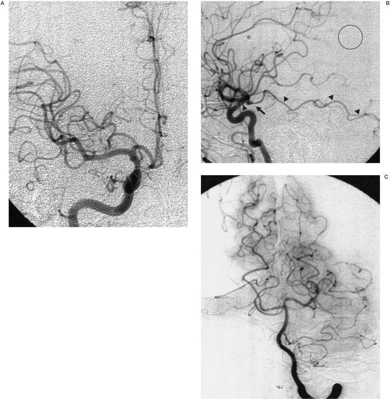
The anterior choroidal artery has the cortical branches to the temporal, parietal, and occipital lobes in the early embryological stage, which later become the posterior cerebral artery distal to the posterior communicating artery (P2-4). Acute embolic stroke occurred in a 57-year-old man with an anterior choroidal artery having such a persistent embryonic branch to the temporal lobe. Recognition of this embryological form of the anterior choroidal artery is clinically important in acute cerebral ischaemia because the cerebral region between the territories supplied by the middle cerebral artery and the anterior choroidal artery is shown on carotid angiography as an avascular area, which could be misunderstood as a region of the acute ischaemia.
Figures
Similar articles
-
[A cardioembolic stroke case involving both the posterior cerebral artery and anterior choroidal artery territories].Rinsho Shinkeigaku. 2007 May;47(5):237-9. Rinsho Shinkeigaku. 2007. PMID: 17585608 Japanese.
-
Fetal-Type Variants of the Posterior Cerebral Artery and Concurrent Infarction in the Major Arterial Territories of the Cerebral Hemisphere.J Investig Med High Impact Case Rep. 2016 Sep 13;4(3):2324709616665409. doi: 10.1177/2324709616665409. eCollection 2016 Jul-Sep. J Investig Med High Impact Case Rep. 2016. PMID: 27660767 Free PMC article.
-
Angiography and treatment of a pial occipital malformation supplied by a posterior temporo-occipital branch of the anterior choroidal artery.Neuroradiology. 1996 May;38 Suppl 1:S157-9. doi: 10.1007/BF02278146. Neuroradiology. 1996. PMID: 8811704
-
[Cerebral deep vascular architectures and subcortical infarcts].Rinsho Shinkeigaku. 2020 Jun 6;60(6):397-406. doi: 10.5692/clinicalneurol.60.cn-001408. Epub 2020 May 19. Rinsho Shinkeigaku. 2020. PMID: 32435049 Review. Japanese.
-
Distal anterior choroidal artery aneurysm associated with an arteriovenous malformation. Intraoperative localization and treatment.Surg Neurol. 2000 Jun;53(6):546-51. doi: 10.1016/s0090-3019(00)00235-4. Surg Neurol. 2000. PMID: 10940421 Review.
Cited by
-
Coexistence of anterior choroidal artery and posterior cerebral artery retia mirabilia presenting with subarachnoid hemorrhage: illustrative case.J Neurosurg Case Lessons. 2024 Jan 1;7(1):CASE23580. doi: 10.3171/CASE23580. Print 2024 Jan 1. J Neurosurg Case Lessons. 2024. PMID: 38163352 Free PMC article.
-
An anomalous hyperplastic anterior choroidal artery associated with an unruptured internal carotid-posterior communicating artery aneurysm.Radiol Case Rep. 2022 Mar 25;17(5):1745-1749. doi: 10.1016/j.radcr.2022.02.071. eCollection 2022 May. Radiol Case Rep. 2022. PMID: 35360188 Free PMC article.
-
Telencephalic variant of the anterior choroidal artery: clinical significance and management considerations.Quant Imaging Med Surg. 2025 Aug 1;15(8):7663-7666. doi: 10.21037/qims-2025-211. Epub 2025 Jul 9. Quant Imaging Med Surg. 2025. PMID: 40785893 Free PMC article. No abstract available.
-
Internal carotid artery-persistent primitive anterior choroidal artery aneurysms: report of two cases and literature review.Acta Neurochir (Wien). 2024 Feb 20;166(1):94. doi: 10.1007/s00701-024-05988-1. Acta Neurochir (Wien). 2024. PMID: 38376611 Review.
References
-
- Abbie AA. The clinical significance of the anterior choroidal artery. Brain. 1933;56:234–246.
-
- Lasjaunias P, Berenstein A, Ter Brugge KG. Surgical Neuroangiography. Vol. 1. Berlin: Springer-Verlag; 2001. Intradural arteries, Clinical vascular anatomy and variations; pp. 479–629.
-
- Herman LH, Fernando OU, Gurdjian ES. The anterior choroidal artery: An anatomical study of its area of distribution. Anat Rec. 1966;154:95–102. - PubMed
-
- Rhoton AL, Fujii K, Fradd B. Microsurgical anatomy of the anterior choroidal artery. Surg Neurol. 1979;12:171–187. - PubMed
LinkOut - more resources
Full Text Sources
