

5-year Angiographic and Clinical Follow-up of Coil-embolised Intradural Saccular Aneurysms. A Single Center Experience
- PMID: 20594497
- PMCID: PMC3572492
- DOI: 10.1177/159101990200800405
5-year Angiographic and Clinical Follow-up of Coil-embolised Intradural Saccular Aneurysms. A Single Center Experience
Abstract
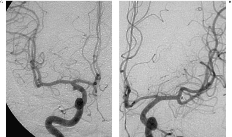
The purpose of the paper is the follow-up of embolised intradural saccular Arterial Aneurysms (AA), excluding giant, dissecting, inflammatory, fusiform or AA associated to BVAM. Since its introduction in 1991, the Guglielmi Detachable Coil has offered protection against aneurysmal rebleeding in the critical few days and months after SAH regardless of the grade. A number of questions remain: is complete angiographic obliteration necessary at first embolisation? What duration of clinical / angiographic follow-up (FU) is required to ensure the risk of haemorrhage has been eliminated? What is the long-term protection against rebleeding? One hundred and two patients with 160 intradural saccular AA embolised before april 1997 were selected for this study. They had at least 5-yrs clinical FU, of which 22 patients had a mid- term (3 years) and 45 patients had a 5-year or more angiographic FU (mean 67,7 months per patient). Twenty-eight embolised AAs with 100% occlusion at 1 year, remained unchanged on the 5-year angiograms. A further 14 patients with complete occlusion at 1 year showed persisting complete occlusion on angiogram at 3-years FU, which in our series means that complete occlusion after the first year post-embolisation implies that the aneurysm will remain completely occluded. All secondary spontaneous thromboses (27.6% of cases), occurred during the first year pos- embolisation. In six patients with subtotal or partial occlusion no change was seen for three consecutive years of FU; none showed later change at 5-year angiography. Below 80% occlusion our series does not provide enough information but we consider the situation instable. No mortality related to the procedure was observed in the unruptured AA group.No bleeding or re-bleeding has occurred since the beginning of our experience (1993) in saccular AA treated by GDC-Coil. Coil-embolisation of properly selected patients is effective in protecting against bleeding or re-bleeding at short and long-term with stable morphological results provided a strict follow-up control is established at short term.
Figures


Similar articles
-
Coil embolization for intracranial aneurysms: an evidence-based analysis.Ont Health Technol Assess Ser. 2006;6(1):1-114. Epub 2006 Jan 1. Ont Health Technol Assess Ser. 2006. PMID: 23074479 Free PMC article.
-
Intradural saccular aneurysms treated by Guglielmi detachable bare coils at a single institution between 1993 and 2005: clinical long-term follow-up for a total of 1810 patient-years in relation to morphological treatment results.Stroke. 2008 Aug;39(8):2288-97. doi: 10.1161/STROKEAHA.107.508234. Epub 2008 Jun 26. Stroke. 2008. PMID: 18583562
-
Treatment of basilar artery bifurcation aneurysms by using Guglielmi detachable coils: a 6-year experience.J Neurosurg. 1999 May;90(5):843-52. doi: 10.3171/jns.1999.90.5.0843. J Neurosurg. 1999. PMID: 10223449
-
Selection of cerebral aneurysms for treatment using Guglielmi detachable coils: the preliminary University of Illinois at Chicago experience.Neurosurgery. 1998 Dec;43(6):1281-95; discussion 1296-7. doi: 10.1097/00006123-199812000-00011. Neurosurgery. 1998. PMID: 9848841 Review.
-
Follow-up angiography of intracranial aneurysms treated with endovascular placement of Guglielmi detachable coils.Neurosurgery. 2002 Feb;50(2):239-49; discussion 249-50. doi: 10.1097/00006123-200202000-00003. Neurosurgery. 2002. PMID: 11844258 Review.
Cited by
-
Periprocedural morbidity and mortality by endovascular treatment of cerebral aneurysms with GDC: a retrospective 12-year experience of a single center.Neurosurg Rev. 2007 Apr;30(2):117-25; discussion 125-6. doi: 10.1007/s10143-006-0059-z. Epub 2007 Jan 11. Neurosurg Rev. 2007. PMID: 17216530
-
Recurrent or new symptomatic cerebral aneurysm after previous treatment.Interv Neuroradiol. 2005 Dec 20;11(4):341-8. doi: 10.1177/159101990501100406. Epub 2006 Feb 10. Interv Neuroradiol. 2005. PMID: 20584446 Free PMC article.
-
Coil embolization for intracranial aneurysms: an evidence-based analysis.Ont Health Technol Assess Ser. 2006;6(1):1-114. Epub 2006 Jan 1. Ont Health Technol Assess Ser. 2006. PMID: 23074479 Free PMC article.
References
-
- Yasargil MG. MicroNeurosurgery. Vol. 1. Stuttgart: Thieme; 1984. Microsurgical Anatomy of the Basal Cisterns and Vessels of the Brain, Diagnostic Studies, General Operative Techniques ans Pathological Considerations of the intracranial Aneurysms.
-
- Guglielmi G, Viñuela F, et al. Elethrombosis of saccular aneurysms via endovascular approach. Electrochemical basis, technique and experimental results. J Neurosurgery. 1991;75(part 1):1–7. - PubMed
-
- Guglielmi G, Viñuela F, et al. Electrothrombosis of saccular aneurysm via endovascular approach. Preliminar clinical experience. Neurosurgery. 1991;75(part 2):8–14. - PubMed
-
- Ausman JI. The future of neurovascular surgery. Part I: Intracranial aneurysms. Surg Neurol. 1997;48:98–100. - PubMed
-
- Molyneux A. International Subarachnoid Aneurysms Trial (ISAT) of neurosurgical clipping versus endovascular coiling in 2143 patients with ruptured intracranial aneurysms: a randomised trial. Lancet. 2002;360:1267–1274. - PubMed
LinkOut - more resources
Full Text Sources
Miscellaneous
