

doi: 10.1177/159101990200800102.
Epub 2004 Oct 20.
Clinical Presentation, Imaging and Treatment of Cerebral Venous Thrombosis (CVT)
Affiliations
- PMID: 20594505
- PMCID: PMC3572523
- DOI: 10.1177/159101990200800102
Item in Clipboard
Clinical Presentation, Imaging and Treatment of Cerebral Venous Thrombosis (CVT)
Interv Neuroradiol.
.
No abstract available
Figures
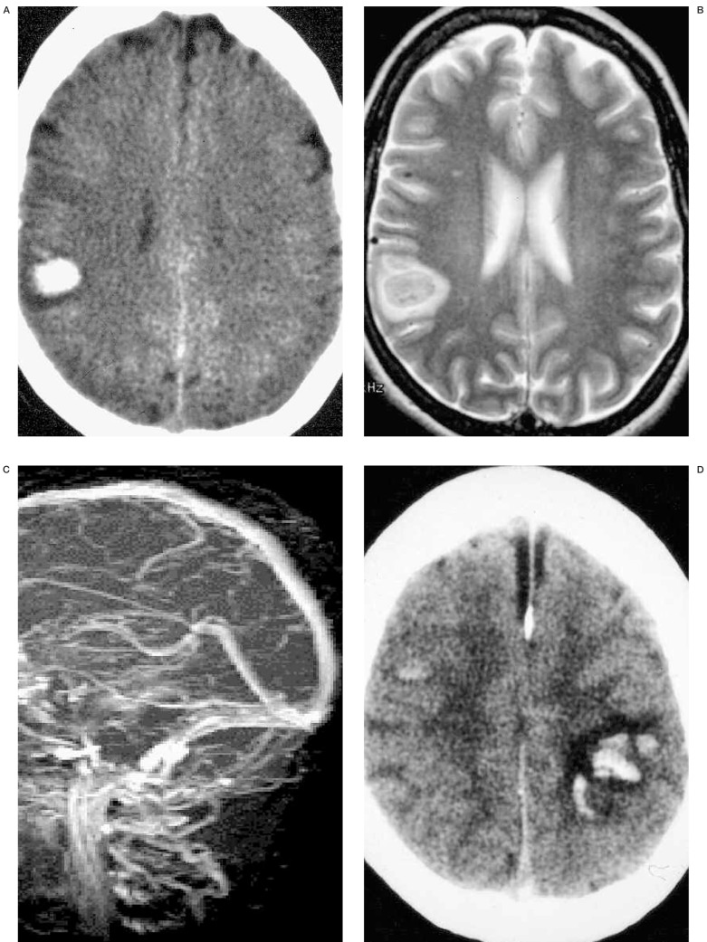
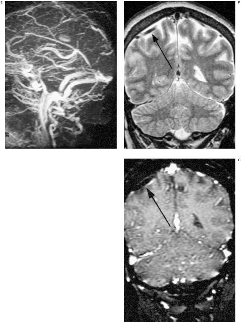
A 32-year-old woman developed generalized seizure at postpartum 2 weeks. Initial non-enhanced brain CT (A) reveals focal right parietal intracranial hemorrhage. T2 weighted axial MR image (B) shows right parietal subacute hematoma and MR venogram (C) does not demonstrate dural sinus occlusion. Because there was no radiologic evidence of CVT, the patient was not given any treatment for 1 week. Follow-up non-enhanced CT (D) shows bilateral intracerebral hemorrhage, dense superior sagittal sinus and subdural fluid collections. MRV (E) demonstrates total occlusion of the superior sagittal sinus. Partial occlusion of distal straight sinus and mid-portion of right transverse sinus are also revealed. Retrograde evaluation of initial MRI (T2 weighted coronal) (F) and MRV source image (G) shows right parietal cortical vein thrombosis without dural sinus involvement. On T2 weighted coronal image (F), thrombosed cortical vein (arrow) shows low signal intensity, so it is difficult to differentiate it from normal vascular signal voids. However, on MRV source image (G), it turns to high signal intensity (arrow) suggesting acute thrombosed cortical vein.
A 32-year-old woman developed generalized seizure at postpartum 2 weeks. Initial non-enhanced brain CT (A) reveals focal right parietal intracranial hemorrhage. T2 weighted axial MR image (B) shows right parietal subacute hematoma and MR venogram (C) does not demonstrate dural sinus occlusion. Because there was no radiologic evidence of CVT, the patient was not given any treatment for 1 week. Follow-up non-enhanced CT (D) shows bilateral intracerebral hemorrhage, dense superior sagittal sinus and subdural fluid collections. MRV (E) demonstrates total occlusion of the superior sagittal sinus. Partial occlusion of distal straight sinus and mid-portion of right transverse sinus are also revealed. Retrograde evaluation of initial MRI (T2 weighted coronal) (F) and MRV source image (G) shows right parietal cortical vein thrombosis without dural sinus involvement. On T2 weighted coronal image (F), thrombosed cortical vein (arrow) shows low signal intensity, so it is difficult to differentiate it from normal vascular signal voids. However, on MRV source image (G), it turns to high signal intensity (arrow) suggesting acute thrombosed cortical vein.
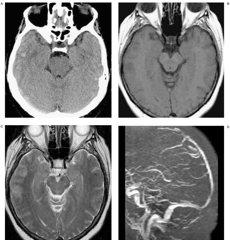
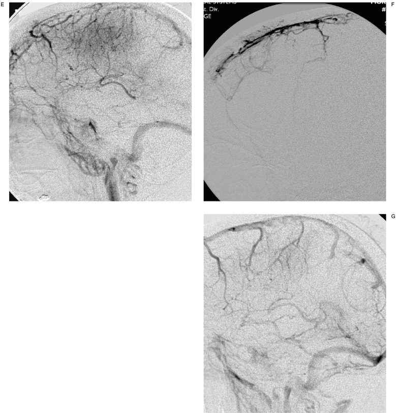
A 62-year-old woman with acute lymphocytic leukemia was treated with heparin due to dural sinus thrombosis. However, the patient's consciousness level deteriorated despite anticoagulation (INR=1.28). Initial non-enhanced CT (A) shows subtle increased attenuation (arrow) on right temporal lobe and right transverse sinus suggesting parenchymal hemorrhage and dural sinus thrombosis, respectively. T2 (B) weighted axial MR demonstrate sulcal effacement and acute parenchymal hemorrhage (arrow) on right temporal lobe. MR venography (C) and venous phase of right internal carotid angiogram (D) reveal nonvisualization of proximal to mid portion of superior sagittal sinus (SSS) and straight sinus. Note there is a lack of parenchymal staining on occipital and temporal lobes on right internal carotid angiogram. Superselective SSS angiography (E) show multifocal filling defects suggesting sinus thrombosis. After infusion of 30mg of t-PA, right internal carotid angiography (F) demonstrates complete re-canalization of SSS and partial recanalization of straight sinus. Three months follow-up MR venography (G) shows patent SSS and straight sinus.
A 62-year-old woman with acute lymphocytic leukemia was treated with heparin due to dural sinus thrombosis. However, the patient's consciousness level deteriorated despite anticoagulation (INR=1.28). Initial non-enhanced CT (A) shows subtle increased attenuation (arrow) on right temporal lobe and right transverse sinus suggesting parenchymal hemorrhage and dural sinus thrombosis, respectively. T2 (B) weighted axial MR demonstrate sulcal effacement and acute parenchymal hemorrhage (arrow) on right temporal lobe. MR venography (C) and venous phase of right internal carotid angiogram (D) reveal nonvisualization of proximal to mid portion of superior sagittal sinus (SSS) and straight sinus. Note there is a lack of parenchymal staining on occipital and temporal lobes on right internal carotid angiogram. Superselective SSS angiography (E) show multifocal filling defects suggesting sinus thrombosis. After infusion of 30mg of t-PA, right internal carotid angiography (F) demonstrates complete re-canalization of SSS and partial recanalization of straight sinus. Three months follow-up MR venography (G) shows patent SSS and straight sinus.
Similar articles
-
Cerebral venous thrombosis: state of the art diagnosis and management.Neuroradiology. 2018 Jul;60(7):669-685. doi: 10.1007/s00234-018-2032-2. Epub 2018 May 11. Neuroradiology. 2018. PMID: 29752489 Review.
-
Cerebral venous thrombosis in Argentina: clinical presentation, predisposing factors, outcomes and literature review.J Stroke Cerebrovasc Dis. 2020 Oct;29(10):105145. doi: 10.1016/j.jstrokecerebrovasdis.2020.105145. Epub 2020 Jul 28. J Stroke Cerebrovasc Dis. 2020. PMID: 32912503 Review.
-
Pediatric Cortical Vein Thrombosis: Frequency and Association With Venous Infarction.Stroke. 2016 Mar;47(3):866-8. doi: 10.1161/STROKEAHA.115.011291. Stroke. 2016. PMID: 26888536 Free PMC article.
-
Cerebral Venous Thrombosis Headache.Curr Pain Headache Rep. 2019 May 30;23(7):47. doi: 10.1007/s11916-019-0786-9. Curr Pain Headache Rep. 2019. PMID: 31147848 Review.
-
Cerebral venous thrombosis--clinical presentations.J Pak Med Assoc. 2006 Nov;56(11):513-6. J Pak Med Assoc. 2006. PMID: 17183979 Review.
Cited by
-
Successful intravascular ultrasound thrombolysis of dural sinus thrombosis with pre-existing subarachnoid and intraparenchymal hemorrhages.Interv Neuroradiol. 2010 Dec;16(4):455-8. doi: 10.1177/159101991001600414. Epub 2010 Dec 17. Interv Neuroradiol. 2010. PMID: 21162778 Free PMC article.
-
Cerebral sinovenous thrombosis. Neuroimaging diagnosis and clinical management.Interv Neuroradiol. 2008 Nov 11;14 Suppl 2(Suppl 2):35-40. doi: 10.1177/15910199080140S208. Epub 2009 Jan 2. Interv Neuroradiol. 2008. PMID: 20557799 Free PMC article.
-
Intracerebral Hemorrhage due to Thrombosis with Thrombocytopenia Syndrome after Vaccination against COVID-19: the First Fatal Case in Korea.J Korean Med Sci. 2021 Aug 9;36(31):e223. doi: 10.3346/jkms.2021.36.e223. J Korean Med Sci. 2021. PMID: 34402235 Free PMC article.
-
Treatment of progressive cerebral sinuses thrombosis with local thrombolysis.Interv Neuroradiol. 2012 Mar;18(1):89-96. doi: 10.1177/159101991201800112. Epub 2012 Mar 16. Interv Neuroradiol. 2012. PMID: 22440606 Free PMC article. Clinical Trial.
-
Bilateral corpus callosum and corona radiata infarction due to cerebral venous sinus thrombosis presenting as headache and acute reversible aphasia: a rare case report.BMC Neurol. 2020 Jun 19;20(1):249. doi: 10.1186/s12883-020-01829-7. BMC Neurol. 2020. PMID: 32560642 Free PMC article.
References
-
- Rosendaal FR. Thrombosis in the young: epidemiology and risk factors. A focus on venous thrombosis. Thromb Haemost. 1997;78:1–6. - PubMed
-
- Manco-Johnson MJ, Nuss R, et al. Combined thrombolytic and anticoagulant therapy for venous thrombosis in children. J Pediatr. 2000;136:446–453. - PubMed
-
- Ameri A, Bousser MG. Cerebral venous thrombosis. Neurol Clin. 1992;10:87–111. - PubMed
-
- de Bruijn SF, Stam J. Randomized, placebo-controlled trial of anticoagulant treatment with low-molecular-weight heparin for cerebral sinus thrombosis. Stroke. 1999;30:484–488. - PubMed
LinkOut - more resources
Full Text Sources
