

doi: 10.1177/159101990200800107.
Epub 2004 Oct 20.
Usefulness of Multidetector 3D-CT Angiography in the Evaluation of Infantile Perimedullary Spinal Arteriovenous Fistula
Affiliations
- PMID: 20594511
- PMCID: PMC3572521
- DOI: 10.1177/159101990200800107
Item in Clipboard
Usefulness of Multidetector 3D-CT Angiography in the Evaluation of Infantile Perimedullary Spinal Arteriovenous Fistula
Interv Neuroradiol.
.
Abstract
We report an infantile huge perimedullary spinal arteriovenous fistula (SAVF) associated with Hereditary-Hemorrhagic-Telangiectasia (HHT), which was treated by glue embolization in one session. Three-dimensional Multidetector Computed Tomography Angiography (3D-MCTA) was useful in pre- and post-endovascular intervention.
Figures
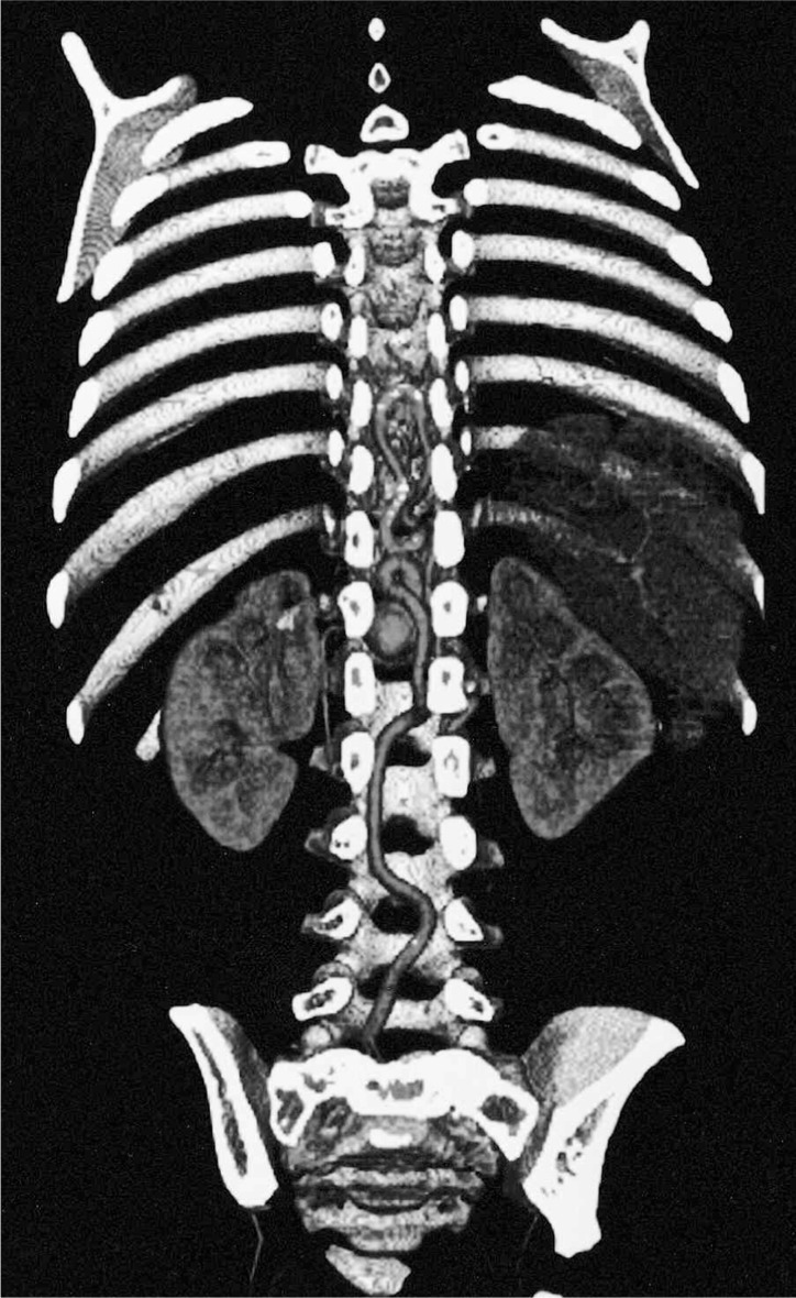
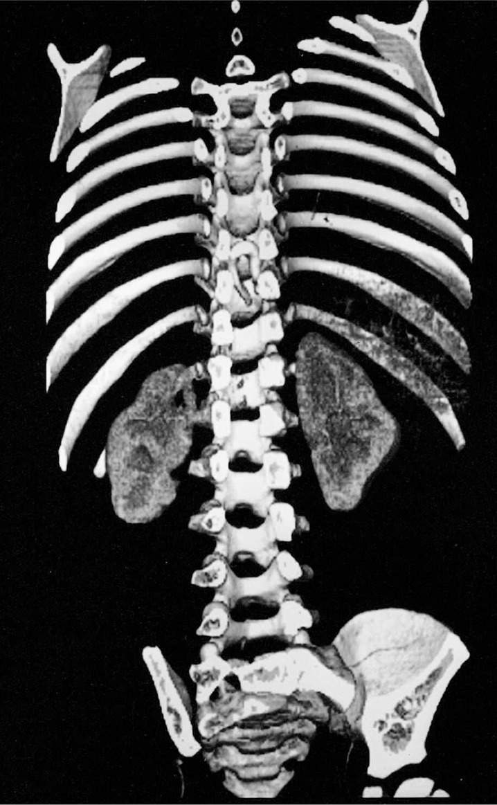
Three dimensional reconstruction arteriography obtained by multidetector helical computed tomography confirm the arteriovenous malformation with the cutting images of the spinal canal.
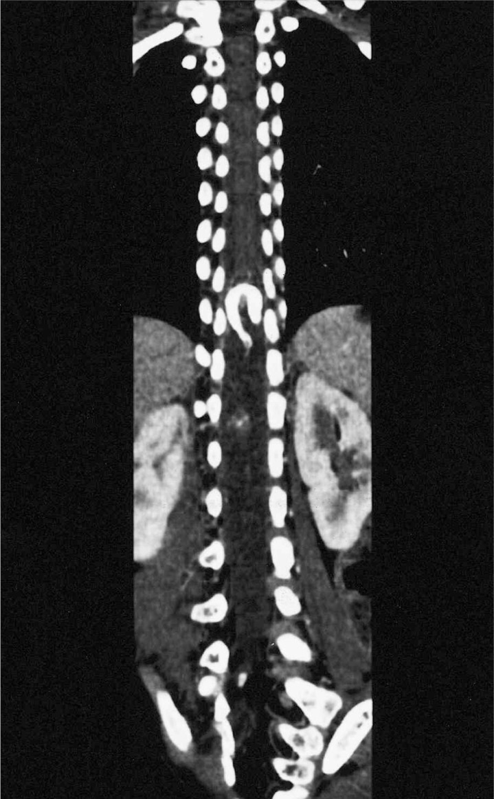
Pre-interventional curved MPR image shows marked dilated vessels of the spinal arteriovenous malformation.
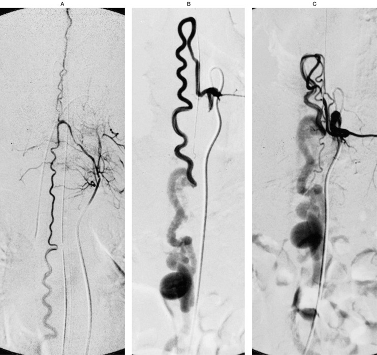
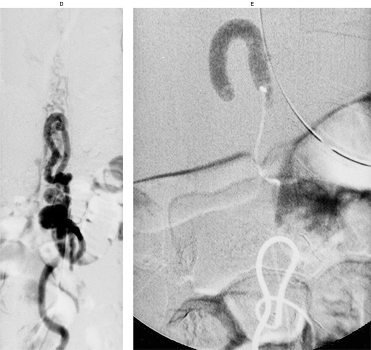
A) Left thyrocervical arteriography demonstrates anterior spinal artery connecting to the radiculo-medullary artery from the left 9 th intercostals artery with a single hole fistula. B) Left 7 th intercostal arteriography shows radiculo-medullary artery connecting to a single hole fistula.at the ventral surface of spinal cord at th elevel Th10. C) Left 9 thintercostal arteriography shows radiculo-medullary artery connecting to a single hole fistula at the ventral surface of spinal cord at th elevel Th10. D) Left 1st. Lumbar arteriography demonstrates radiculopial artery, which is the biggest feeding artery connecting to a single hole fistula with the dilated draining vein. E) Glue embolization closes the shunt between multifeeders and single-hole fistula.
A) Left thyrocervical arteriography demonstrates anterior spinal artery connecting to the radiculo-medullary artery from the left 9 th intercostals artery with a single hole fistula. B) Left 7 th intercostal arteriography shows radiculo-medullary artery connecting to a single hole fistula.at the ventral surface of spinal cord at th elevel Th10. C) Left 9 thintercostal arteriography shows radiculo-medullary artery connecting to a single hole fistula at the ventral surface of spinal cord at th elevel Th10. D) Left 1st. Lumbar arteriography demonstrates radiculopial artery, which is the biggest feeding artery connecting to a single hole fistula with the dilated draining vein. E) Glue embolization closes the shunt between multifeeders and single-hole fistula.
Post-interventional three-dimensional reconstruction arteriography shows glue cast and disappearance of the abnormal shunt.
Post-interventional curved MPR images confirm complete disappearance of SAVF except embolized glue deposition.
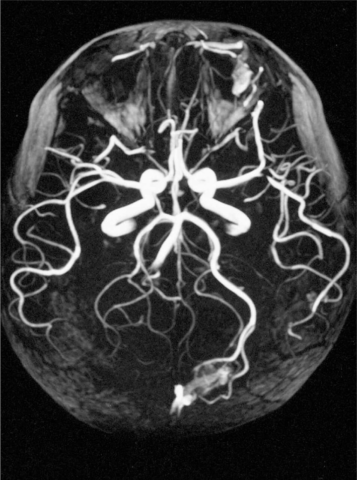
Magnetic resonance angiography demonstrate asymptomatic multiple cortical small AVM.
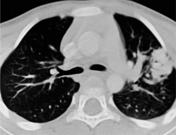
Helical compeuted tomography show pulmonary AVMs at left segment 3.
Similar articles
-
Giant spinal perimedullary fistula in hereditary haemorrhagic telangiectasia: diagnosis, endovascular treatment and review of the literature.Neuroradiology. 2003 Nov;45(11):830-6. doi: 10.1007/s00234-003-1044-7. Epub 2003 Oct 14. Neuroradiology. 2003. PMID: 14557903 Review.
-
Acute thrombosis of a giant perimedullary arteriovenous fistula in a pediatric HHT patient.Interv Neuroradiol. 2022 Apr;28(2):132-135. doi: 10.1177/15910199211022499. Epub 2021 May 29. Interv Neuroradiol. 2022. PMID: 34053318 Free PMC article.
-
Endovascular treatment of spinal arteriovenous fistula in a young child with hereditary hemorrhagic telangiectasia. Case report.J Neurosurg. 2005 Nov;103(5 Suppl):462-5. doi: 10.3171/ped.2005.103.5.0462. J Neurosurg. 2005. PMID: 16302622
-
Spinal cord arteriovenous malformations in two patients with hereditary hemorrhagic telangiectasia.Childs Nerv Syst. 1999 Mar;15(2-3):80-3. doi: 10.1007/s003810050336. Childs Nerv Syst. 1999. PMID: 10230660
-
Spinal giant intradural perimedullary arteriovenous fistula: clinical and neuroradiological study in one case with review of literature.Surg Neurol. 1996 Jun;45(6):524-31; discussion 531-2. doi: 10.1016/0090-3019(95)00433-5. Surg Neurol. 1996. PMID: 8638237 Review.
Cited by
-
Pediatric high-flow, cervical spinal, macro-arteriovenous fistula, treated with the endovascular cotton candy glue injection technique.Childs Nerv Syst. 2010 Nov;26(11):1633-8. doi: 10.1007/s00381-010-1181-3. Epub 2010 Jun 3. Childs Nerv Syst. 2010. PMID: 20521056
References
-
- Terae K, Miyasaka K, et al. Three-dimensional reconstructed CT angiography. Detection of the Adamkiewicz artery. Abstract; 30th Japanese Neuroradiological congress; 18-22 March, 2006; Osaka, Japan. 2001.
-
- Lasjaunias P, TerBrugge K. Spinal Arteriovenous Malformation. Vascular Disease in Neonates, Infants and Children. New York; Tokyo: Springer Berlin Heidelberg; 1997. pp. 565–591.
-
- Berenstein A, Lasjaunias P. Surgical Neuroangiography. Vol. 5. Berlin Heidelberg; New York; Tokyo: Springer; 1992. Endovascular treatment of spine and spinal cord lesions.
-
- Halbach VV, Higashida R, et al. Treatment of giant intradural perimedullary arteriovenous fistulas. Neurosurgery. 1993;33(6):972–984. - PubMed
-
- Rodesch G, Pongpech S, et al. Spinal Cord Arteriovenous Malformations in a Pediatric Population of Children below 15 Years of Age. The place of Endovascular management. Interventional Neuroradiology. 1995;1:29–42. - PubMed
LinkOut - more resources
Full Text Sources
