

Multiple dural arteriovenous fistulas. Radiologic progression and endovascular cure. Case report
- PMID: 20594527
- PMCID: PMC3576611
- DOI: 10.1177/159101990200800210
Multiple dural arteriovenous fistulas. Radiologic progression and endovascular cure. Case report
Abstract
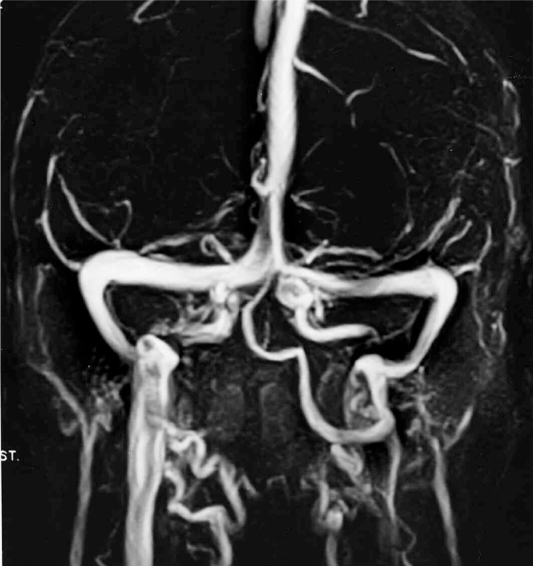
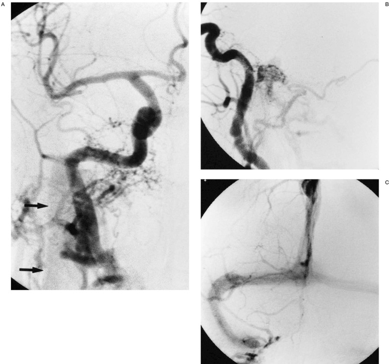
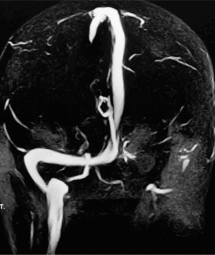
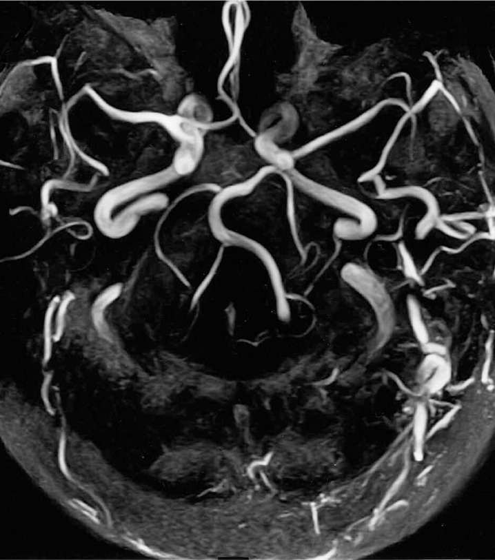
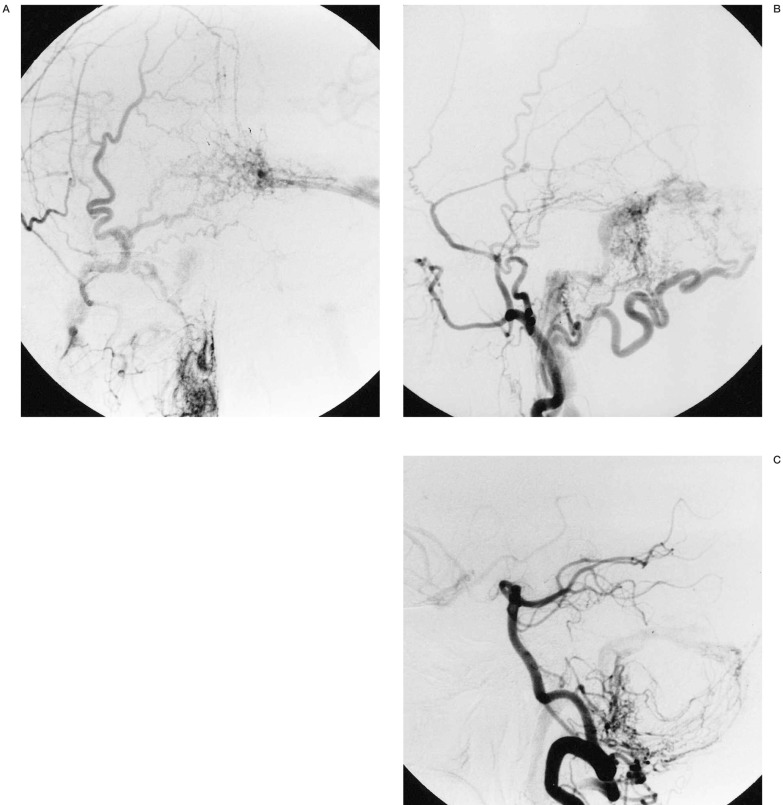
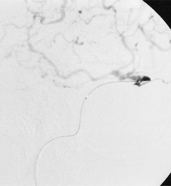
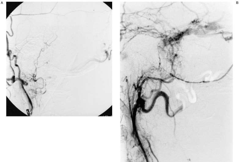
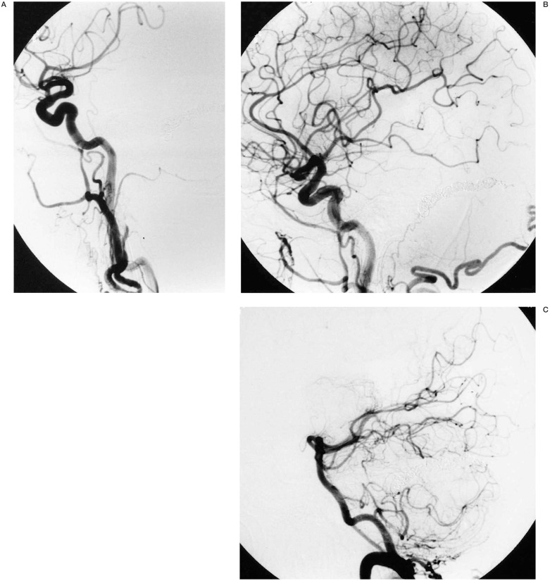
Dural arteriovenous fistulas are most probably acquired lesions. However, they have been rarely encountered de novo.We present a unique case of a 71-year-old woman who initially presented with right-sided dural arteriovenous fistula (DAVF), which spontaneously resolved after diagnostic arteriography. She later developed asymptomatic occlusion of the left transverse sinus. Five years after her initial presentation she developed left-sided pulse-synchronous tinnitus. MRA and catheter angiography showed a complex type IV DAVF between the left transverse sinus and multiple dural branches arising from both left and right external carotid arteries. The left transverse sinus was isolated from the torcula herophili, with stenosis of the sigmoid sinus. Extensive cortical venous drainage was demonstrated. Endovascular cure was effected by polyvinyl alcohol particle and absolute alcohol occlusion of the dominant dural supply, and transvenous coil occlusion of the left transverse sinus. The patient's symptoms resolved almost immediately. This unique case demonstrates that dural sinus occlusion and DAVFs may co-exist, but there may not be a causal relationship. It is likely that both DAVFs and sinus occlusion are manifestations of the same disease process characterised by a pro-thrombotic state and secondary angiogenesis. It is important to recognise that changes in symptomatology, even long after apparent disappearance of a lesion may indicate recurrence, and careful follow up is advocated.
Figures
Similar articles
-
Endovascular Recanalization of Occluded Dural Sinus in Patient with Dural Arteriovenous Fistulas: Case Report and Literature Review.World Neurosurg. 2018 Jun;114:269-273. doi: 10.1016/j.wneu.2018.03.140. Epub 2018 Mar 27. World Neurosurg. 2018. PMID: 29602009 Review.
-
[Evolution of angiographic signs of venous hypertension and clinical signs of intracranial hypertension in intracranial dural arteriovenous fistulas].J Neuroradiol. 1999 Mar;26(1):49-58. J Neuroradiol. 1999. PMID: 10363442 French.
-
Occurrence of De Novo Dural Arteriovenous Fistula after Transvenous Embolization of Dural Arteriovenous Fistula : Case Reports of Two Patients.J Korean Neurosurg Soc. 2022 Jul;65(4):598-602. doi: 10.3340/jkns.2021.0144. Epub 2022 Apr 14. J Korean Neurosurg Soc. 2022. PMID: 35418004 Free PMC article.
-
[Multiple dural arteriovenous fistulas involving both the cavernous sinus and the posterior fossa: report of two cases and review of the literature].No Shinkei Geka. 2001 Nov;29(11):1065-72. No Shinkei Geka. 2001. PMID: 11758314 Review. Japanese.
-
[A Case of Cavernous Sinus Dural Arteriovenous Fistula after Transverse-Sigmoid Sinus Dural Arteriovenous Fistula Treatment with Radical Transvenous Coil Embolization].No Shinkei Geka. 2015 Aug;43(8):753-7. doi: 10.11477/mf.1436203114. No Shinkei Geka. 2015. PMID: 26224471 Japanese.
Cited by
-
Cerebral venous sinus thrombosis and dural arteriovenous fistula associated with protein S deficiency: a case series study.BMC Neurol. 2022 May 2;22(1):164. doi: 10.1186/s12883-022-02693-3. BMC Neurol. 2022. PMID: 35501720 Free PMC article.
References
-
- Houser OW, Campbell JK, et al. Arteriovenous malformation affecting the transverse dural venous sinus-an acquired lesion. Mayo Clin Proced. 1979;54:651–661. - PubMed
-
- Vinuela F, Fox AJ, et al. Spontaneous carotid-cavernous fistulas: clinical, radiological and therapeutic considerations. J Neurosurg. 1984;60:976–984. - PubMed
-
- Wakamoto H, Miyazaki H, et al. The natural history of a dural arteriovenous fistula associated with sinus thrombosis: a case report. No Shinkei Geka. 1999;27:563–568. - PubMed
LinkOut - more resources
Full Text Sources
