

Spontaneous Thrombosis of the Pseudoaneurysm of Right SCA after an Attempt at Embolisation. A Case Report
- PMID: 20594531
- PMCID: PMC3576615
- DOI: 10.1177/159101990200800214
Spontaneous Thrombosis of the Pseudoaneurysm of Right SCA after an Attempt at Embolisation. A Case Report
Abstract
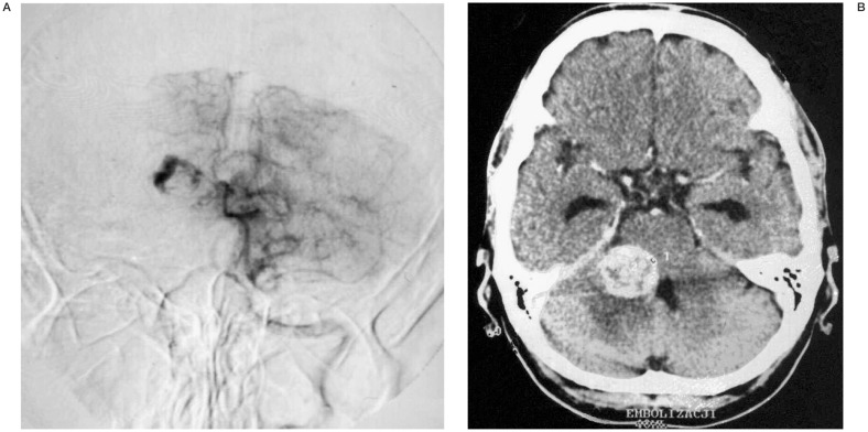
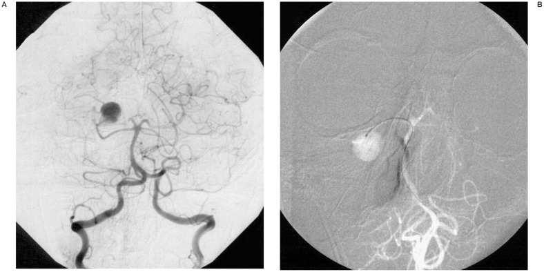
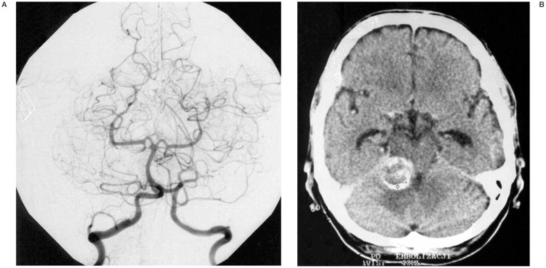
Spontaneous thrombosis of intracranial aneurysms rare, mostly affecting giant aneurysms with narrow necks. We present the case of 34 y/o man with pseudoaneurysm that developed in the course of SAH. The initial CT scan showed an isolated, well-defined hematoma within the right cerebellar hemisphere, digital subtraction angiogram (DSA) performed in a regional hospital showed an irregular shaped aneurysm of the distal segment of the right SCA. The patient was sent to our department, where diagnostic DSA, performed before embolisation revealed an entirely different morphology of the aneurysm. It became larger, round and no other functional branches distal to it were found (picture of "a balloon on a string"). During supraselective catheterization, when microcatheter and microguidewire were already in the right SCA a technical problem of our angio-machine occurred, so the intervention had to be postponed. A week later, a second attempt at embolisation was made. This time an initial DSA showed a lack of filling of the aneurysm sac and thrombosis of the main trunk of the right SCA. The patient remained clinically stable. He was discharged from our hospital five days later.
Figures
Similar articles
-
Spontaneous total thrombosis of distal superior cerebellar artery aneurysm.Acta Neurochir (Wien). 2001 Aug;143(8):837-42; discussion 842-3. doi: 10.1007/s007010170039. Acta Neurochir (Wien). 2001. PMID: 11678406 Review.
-
Multiple aneurysms of the distal posterior inferior cerebellar artery: two case reports.Minim Invasive Neurosurg. 2008 Oct;51(5):249-52. doi: 10.1055/s-0028-1082302. Epub 2008 Oct 14. Minim Invasive Neurosurg. 2008. PMID: 18855286
-
Spontaneous thrombosis of an intracranial giant aneurysm.Interv Neuroradiol. 1999 Dec 20;5(4):327-32. doi: 10.1177/159101999900500410. Epub 2001 May 15. Interv Neuroradiol. 1999. PMID: 20670531
-
Endovascular Treatment of Distal Lenticulostriate Artery Aneurysm by Selective Catheterization of Artery with Balloon-Blocking Technique: 2-Dimensional Video Illustration.World Neurosurg. 2020 Apr;136:220. doi: 10.1016/j.wneu.2020.01.054. Epub 2020 Jan 16. World Neurosurg. 2020. PMID: 31954888
-
Ruptured Distal Posterior Inferior Cerebellar Artery (PICA) Aneurysms Associated with Cerebellar Arterial Venous Malformations (AVMs): A Case Series and Review of the Literature Demonstrating the Need for Angiographic Evaluation and Feasibility of Endovascular Treatment.World Neurosurg. 2017 Jan;97:751.e7-751.e13. doi: 10.1016/j.wneu.2016.10.081. Epub 2016 Oct 25. World Neurosurg. 2017. PMID: 27793767 Review.
Cited by
-
Thrombosis and recanalization of small saccular cerebral aneurysm : two case reports and a suggestion for possible mechanism.J Korean Neurosurg Soc. 2014 May;55(5):280-3. doi: 10.3340/jkns.2014.55.5.280. Epub 2014 May 31. J Korean Neurosurg Soc. 2014. PMID: 25132936 Free PMC article.
-
Delayed presentation and rupture of an intracranial pseudoaneurysm following penetrating trauma: illustrative case.J Neurosurg Case Lessons. 2025 Mar 10;9(10):CASE24745. doi: 10.3171/CASE24745. Print 2025 Mar 10. J Neurosurg Case Lessons. 2025. PMID: 40064009 Free PMC article.
-
Spontaneous Occluded Anterior Communicating Artery Aneurysm during Coil Embolization Treated with One Coil Insertion into Remaining Stump.J Cerebrovasc Endovasc Neurosurg. 2015 Sep;17(3):246-51. doi: 10.7461/jcen.2015.17.3.246. Epub 2015 Sep 30. J Cerebrovasc Endovasc Neurosurg. 2015. PMID: 26523260 Free PMC article.
References
-
- Rosta L, Battaglia R, et al. Italian co-operative study on giant intracranial aneurysms: Radiological data. Acta Neurochirurgica. 1988;42(suppl):53–59. - PubMed
-
- Whittle IR, Williams DB, et al. Spontaneous thrombosis of a giant intracranial aneurysm and ipsilateral internal carotid artery. J Neurosurg. 1982;56:287–289. - PubMed
-
- Gerber DD, Sahel M, et al. Complete spontaneous thrombosis of a giant intracranial aneurysm. Neuroradiology. 1994;36:316–317. - PubMed
-
- Lyall A. Large aneurysm of the circle of Willis with spontaneous cure by thrombosis. Br J Med. 1936;2:282.
-
- Katayama Y, Tsubokawa T, et al. Growth of totally thrombosed giant aneurysm within the posterior cranial fossa. Neuroradiology. 1991;33:168–170. - PubMed
LinkOut - more resources
Full Text Sources
