. 2010 Nov;95(11):1823-31.
doi: 10.3324/haematol.2010.025015. Epub 2010 Jun 30.

Macrophages as novel target cells for erythropoietin

Affiliations

Macrophages as novel target cells for erythropoietin

Lilach Lifshitz et al. Haematologica. 2010 Nov.

Abstract

Background: Our original demonstration of immunomodulatory effects of erythropoietin in multiple myeloma led us to the search for the cells in the immune system that are direct targets for erythropoietin. The finding that lymphocytes do not express erythropoietin receptors led to the hypothesis that other cells act as direct targets and thus mediate the effects of erythropoietin. The finding that erythropoietin has effects on dendritic cells thus led to the question of whether macrophages act as target cells for erythropoietin.

Design and methods: The effects of erythropoietin on macrophages were investigated both in-vivo and in-vitro. The in-vivo studies were performed on splenic macrophages and inflammatory peritoneal macrophages, comparing recombinant human erythropoietin-treated and untreated mice, as well as transgenic mice over-expressing human erythropoietin (tg6) and their control wild-type counterparts. The in-vitro effects of erythropoietin on macrophage surface markers and function were investigated in murine bone marrow-derived macrophages treated with recombinant human erythropoietin.

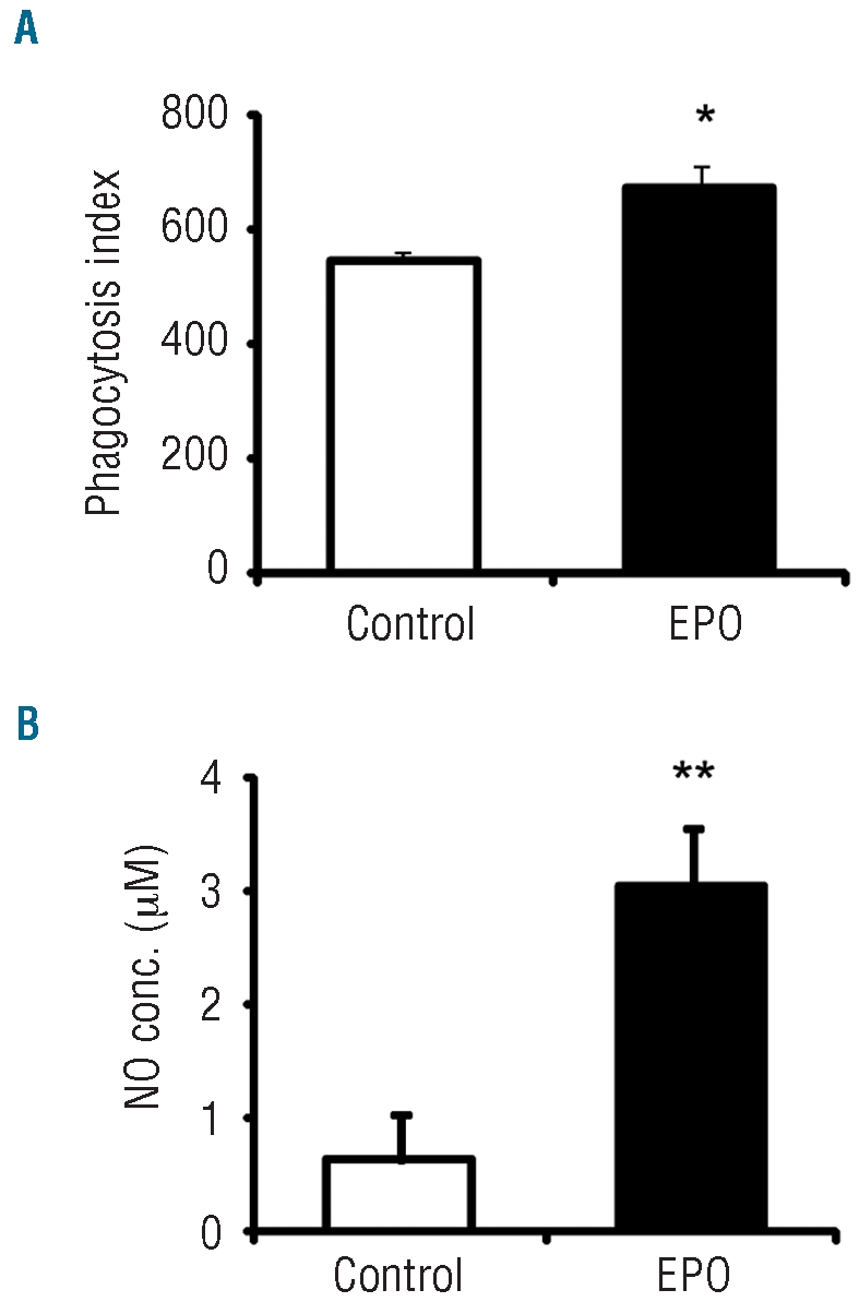
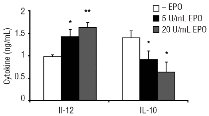
Results: Erythropoietin was found to have effects on macrophages in both the in-vivo and in-vitro experiments. In-vivo treatment led to increased numbers of splenic macrophages, and of the splenic macrophages expressing CD11b, CD80 and major histocompatibility complex class II. The peritoneal inflammatory macrophages obtained from erythropoietin-treated mice displayed increased expression of F4/80, CD11b, CD80 and major histocompatibility complex class II, and augmented phagocytic activity. The macrophages derived in-vitro from bone marrow cells expressed erythropoietin receptor transcripts, and in-vitro stimulation with erythropoietin activated multiple signaling pathways, including signal transducer and activator of transcription (STAT)1 and 5, mitogen-activated protein kinase, phosphatidylinositol 3-kinase and nuclear factor kappa B. In-vitro erythropoietin treatment of these cells up-regulated their surface expression of CD11b, F4/80 and CD80, enhanced their phagocytic activity and nitric oxide secretion, and also led to augmented interleukin 12 secretion and decreased interleukin 10 secretion in response to lipopolysaccharide.

Conclusions: Our results show that macrophages are direct targets of erythropoietin and that erythropoietin treatment enhances the pro-inflammatory activity and function of these cells. These findings point to a multifunctional role of erythropoietin and its potential clinical applications as an immunomodulating agent.


Figures

Figure 1.
Figure 1.
In-vivo effects of erythropoietin (EPO) manifested in splenic macrophages. Flow cytometry analysis of splenocytes from C57BL/6 mice injected three times (every other day during 1 week) with 180 U rHuEPO (EPO) or with the diluent (DILU) as a negative control, and from tg6 mice and their wild type (wt) littermates. (A) Density plots for F4/80 and MHC class II expression. (B) Summary of the number of splenic macrophages: F4/80+ splenocytes. (C) Summary of the number of macrophages: F4/80+ splenocytes also expressing CD11b and CD80/MHC class II. The graph and table represent the mean ± S.E.M of three independent experiments, each including three female mice per experimental group, *P<0.05, **P<0.01 for EPO/tg6 versus diluent/wt, respectively.
Figure 2.
Figure 2.
In-vivo effects of erythropoietin (EPO) on inflammatory macrophages. Flow cytometry analysis of thioglycollate-induced peritoneal macrophages from C57BL/6 mice injected three times (every other day during 1 week) with 180 U rHuEPO (EPO) or with diluent (DILU) as a negative control, and from tg6 mice and their wild-type (wt) littermates. (A and B) Peritoneal macrophages (identified by CD11b expression) were analyzed for CD11b, F4/80, CD80 and MHC class II cell surface expression. Surface expression of these molecules is represented as black or gray histograms. Isotype controls (Iso) are represented by broken line histograms. Histograms represent one of at least three independent experiments, displaying similar results. (A) Data from rHuEPO-injected mice and diluent injected mice are represented by black and gray histograms, respectively. (B) Data from tg6 mice and their wt littermates are represented by black and gray histograms, respectively. (C) Analysis of thioglycollate-induced peritoneal macrophages incubated with FITC-labeled E. coli bacteria. Phagocytosis index = (% of FITC-positive cells) × (mean fluorescence intensity). *P<0.05 for EPO/tg6 versus diluent/wt, respectively. The figure presents data from a total of three experiments.
Figure 3.
Figure 3.
Erythropoietin receptor (EPO-R) expression and activation in bone marrow-derived macrophages (BMDM). (A) EPO-R expression in BMDM. EPO-R mRNA from murine BMDM (99% purity) was reverse-transcribed and subjected to PCR analysis for murine EPO-R and actin transcripts using oligonucleotide primers, thus yielding 300 and 457 bp fragments, respectively. Lane 1: RNA of BMDM. Lane 2: Positive control. cDNA of BaF/3 cells transfected with murine EPO-R. Lane 3: cDNA of BMDM. Lane 4: Negative control. cDNA of MBA cells. (B and C) Stimulation of BMDM with EPO activates multiple signaling pathways. BMDM were treated in-vitro with 50 U/mL of recombinant human EPO (rHuEPO) for the indicated time periods. (B) Cell lysates of the cytosolic fraction were subjected to immunoblot analysis with the indicated antibodies. STAT5, STAT1 and STAT3 signaling was determined by phosphorylation. Total STAT levels are depicted for normalization. (C) Cell lysates of the cytosolic (ERK and AKT immunoblots) and nuclear fractions (p65 immunoblot) were subjected to immunoblot analysis with the indicated antibodies. MAPK and PI3K signaling was determined by ERK1/2 and AKT phosphorylation, respectively. Total ERK2 and AKT levels are depicted for normalization. The NFκB signaling was detected by nuclear p65 internalization. Nuclear histone is depicted for normalization. All micrographs represent one of at least three similar independent experiments. (D) Neutralization of EPO mediated signaling by anti-human EPO antibody. BMDM were treated in-vitro for 10 min with 50 U/mL rHuEPO in the presence or absence of 10 μg/mL of anti human EPO antibody, or with antibody alone. Cell lysates of the cytosolic fraction were subjected to immunoblot analysis with the indicated antibodies. pSTAT5 and total STAT5 levels are depicted, NT - non treated; Ab - anti-human EPO antibody. Immunoblots represent one of at least three independent experiments, displaying similar results.
Figure 4.
Figure 4.
Erythropoietin (EPO) in-vitro leads to enhanced innate functions of bone marrow-derived macrophages (BMDM). BMDM were cultured in-vitro for 24 h with 5 U/mL rHuEPO (EPO) or without (control). (A) Cells were washed and incubated with FITC-labeled E. coli bacteria, and then subjected to FACS analysis. Phagocytosis index = (% of FITC positive cells) × (mean fluorescence intensity). (B) Supernatants were collected and nitric oxide (NO) secretion was detected using Griess reagent. Graphs represent the mean ± S.E.M of three independent experiments. *P<0.05, **P<0.01 for EPO versus control.
Figure 5.
Figure 5.
Erythropoietin (EPO) in-vitro affects the marrow-derived macrophage (BMDM) phenotype. BMDM were cultured in-vitro with (EPO) or without (control) 5 U/mL rHuEPO for 24 h. Flow cytometry analysis of BMDM of CD11b, F4/80 and CD80. Surface expression of these molecules is represented as black (EPO treated) or gray (control) histograms. Isotype controls (Iso) are represented by broken line histograms. Graphs represent the mean percentage of mean fluorescence intensity (MFI) increase of EPO-treated cells versus control cells of three independent experiments. *P<0.05 for EPO versus control.
Figure 6.
Figure 6.
Erythropoietin (EPO) in-vitro leads to increased secretion of IL-12 and decreased IL-10 secretion by marrow-derived macrophages (BMDM). BMDM were cultured in-vitro with 0.05 mg/mL lipopolysaccharide in the presence or absence of rHuEPO for 24 h. Cell culture supernatants were analyzed for the levels of IL-12 and IL-10, by ELISA. Graphs represent the mean cytokine concentration ± S.E.M. of at least three independent experiments. *P<0.05, **P<0.01 for EPO-treated cells versus non-EPO-treated cells.

Comment in

References

    1. Arcasoy MO. The non-haematopoietic biological effects of erythropoietin. Br J Haematol. 2008;141(1):14–31. - PubMed
    1. Mittelman M, Zeidman A, Fradin Z, Magazanik A, Lewinski UH, Cohen A. Recombinant human erythropoietin in the treatment of multiple myeloma-associated anemia. Acta Haematol. 1997;98(4):204–10. - PubMed
    1. Prutchi-Sagiv S, Golishevsky N, Oster HS, Katz O, Cohen A, Naparstek E, et al. Erythropoietin treatment in advanced multiple myeloma is associated with improved immunological functions: could it be beneficial in early disease? Br J Haematol. 2006;135(5):660–72. - PubMed
    1. Mittelman M, Neumann D, Peled A, Kanter P, Haran-Ghera N. Erythropoietin induces tumor regression and antitumor immune responses in murine myeloma models. Proc Natl Acad Sci USA. 2001;98(9):5181–6. - PMC - PubMed
    1. Katz O, Barzilay E, Skaat A, Herman A, Mittelman M, Neumann D. Erythropoietin induced tumour mass reduction in murine lymphoproliferative models. Acta Haematol. 2005;114(3):177–9. - PubMed

Publication types

MeSH terms