. 2010 Nov 15;182(10):1273-81.
doi: 10.1164/rccm.201001-0001OC. Epub 2010 Jul 1.

Host-derived interleukin-5 promotes adenocarcinoma-induced malignant pleural effusion

Affiliations

Host-derived interleukin-5 promotes adenocarcinoma-induced malignant pleural effusion

Georgios T Stathopoulos et al. Am J Respir Crit Care Med. .

Abstract

Rationale: IL-5 is a T helper 2 cytokine important in the trafficking and survival of eosinophils. Because eosinophils can be found in malignant pleural effusions (MPE) from mice and humans, we asked whether IL-5 is involved in the pathogenesis of MPE.

Objectives: To determine the role of IL-5 in MPE formation.

Methods: The effects of IL-5 on experimental MPE induced in C57BL/6 mice by intrapleural injection of syngeneic lung (Lewis lung cancer [LLC]) or colon (MC38) adenocarcinoma cells were determined using wild-type (il5(+/+)) and IL-5-deficient (il5⁻(/)⁻) mice, exogenous administration of recombinant mouse (rm) IL-5, and in vivo antibody-mediated neutralization of endogenous IL-5. The direct effects of rmIL-5 on LLC cell proliferation and gene expression in vitro were determined by substrate reduction and microarray.

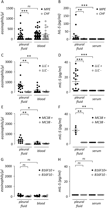
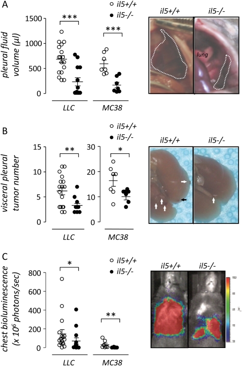
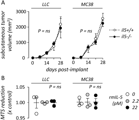
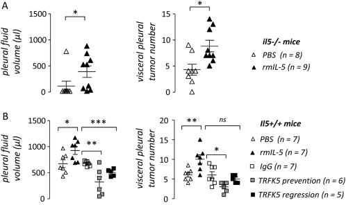
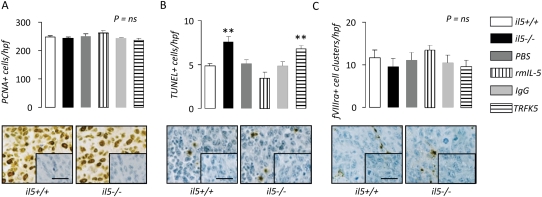
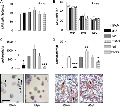
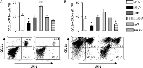
Measurements and main results: Eosinophils and IL-5 were present in human and mouse MPE, but the cytokine was not detected in mouse (LLC) or human (A549) lung and mouse colon (MC38) adenocarcinoma-conditioned medium, suggesting production by host cells in MPE. Compared with il5(+/+) mice, il5⁻(/)⁻ mice showed markedly diminished MPE formation in response to both LLC and MC38 cells. Exogenous IL-5 promoted MPE formation in il5(+/+) and il5⁻(/)⁻ mice, whereas anti-IL-5 antibody treatment limited experimental MPE in il5(+/+) mice. Exogenous IL-5 had no effects on LLC cell proliferation and gene expression; however, IL-5 was found to be responsible for recruitment of eosinophils and tumor-promoting myeloid suppressor cells to MPE in vivo.

Conclusions: Host-derived IL-5 promotes experimental MPE and may be involved in the pathogenesis of human MPE.


Figures

Figure 1.
Figure 1.
Eosinophils and IL-5 in mouse and human malignant pleural effusion (MPE). (A) Pleural fluid and blood eosinophils in humans with pleural effusion caused by malignancy (MPE, n = 55) and heart failure (congestive heart failure [CHF], n = 20). Pleural eosinophils are increased in patients with MPE, compared with CHF. (B) Pleural fluid and serum IL-5 levels in humans with MPE (n = 22) and CHF (n = 16). IL-5 is locally increased in the pleural fluid of patients with MPE. (C) Pleural fluid and blood eosinophil numbers in C57BL/6 mice 14 days after intrapleural delivery of LLC cells (LLC+, n = 17) or phosphate-buffered saline (PBS) control (LLC, n = 8). Pleural eosinophil accumulation is observed after tumor cell injection only. (D) Pleural fluid and serum IL-5 levels in C57BL/6 mice 14 days after intrapleural delivery of LLC cells (LLC+, n = 20) or PBS control (LLC, n = 10). (E) Pleural fluid and blood eosinophil numbers in C57BL/6 mice 11 days after intrapleural delivery of MC38 cells (MC38+, n = 12) or PBS control (MC38, n = 5). Pleural eosinophil accumulation is observed after tumor cell injection only. (F) Pleural fluid and serum IL-5 levels in C57BL/6 mice 11 days after intrapleural delivery of MC38 cells (MC38+, n = 5) or PBS control (MC38, n = 5). (G) Pleural fluid and blood eosinophil numbers in C57BL/6 mice 14 days after intrapleural delivery of B16F10 cells (B16F10+, n = 12) or PBS control (B16F10, n = 8). (H) Pleural fluid and serum IL-5 levels in C57BL/6 mice 14 days after intrapleural delivery of B16F10 cells (B16F10+, n = 5) or PBS control (B16F10, n = 5). Dots = raw data points; lines = mean; bars = SE. *P < 0.05, **P < 0.01, ***P < 0.001. B16F10 = mouse skin melanoma; CHF = congestive heart failure; h = human; LLC = Lewis lung cancer; m = mouse; MC38 = mouse colon adenocarcinoma; MPE = malignant pleural effusion; ns = not significant.
Figure 2.
Figure 2.
IL-5 promotes experimental lung and colon adenocarcinoma–induced malignant pleural effusion (MPE). (A) Pleural fluid volume, (B) pleural tumor number, and (C) bioluminescence of MPEs generated in wild-type (il5+/+, n = 27) and IL-5 knock-out (il5−/−, n = 23) mice on the C57BL/6 background 14 and 11 days after intrapleural delivery of LLC (n = 30) and MC38 (n = 14) cells, respectively. Genetic IL-5 deficiency limits pleural fluid accumulation and intrapleural adenocarcinoma dissemination. Photographs show representative images of MPEs (A, transdiaphragmatic views), of pleural tumors (B, post-lung explantation from thorax; arrows point toward visceral pleural tumors), and of bioluminescence emission (C, day of sacrifice) from il5+/+ and il5−/−mice bearing intrapleural LLC cells. Dots = raw data points; lines = mean; bars = SE. *P < 0.05, ** P < 0.01, ***P < 0.001. LLC = Lewis lung cancer; MC38 = mouse colon adenocarcinoma.
Figure 3.
Figure 3.
IL-5 does not enhance tumor growth in vivo and in vitro. (A) Volume of subcutaneous flank tumors induced in wild-type (il5+/+, n = 10) and IL-5 knock-out (il5−/−, n = 10) mice after subcutaneous injection of 5 × 10⁵ Lewis lung carcinoma (LLC; n = 10) or colon adenocarcinoma (MC38; n = 10) cells. Dots = mean; bars = SE. (B) In vitro cell proliferation rate of LLC and MC38 cells 4 days after exposure to varying concentrations of recombinant mouse IL-5, as determined by a substrate (MTS) reduction assay. Note that 22 pM is the binding constant (kilodalton) of IL-5 for its receptor. Dots = raw data points; lines = mean; bars = SE. LLC = Lewis lung cancer; MC38 = mouse colon adenocarcinoma; ns = not significant.
Figure 4.
Figure 4.
Exogenous IL-5 directly promotes and IL-5 neutralization limits experimental lung adenocarcinoma–induced malignant pleural effusion (MPE). (A) Pleural fluid volume (left) and pleural tumor number (right) of MPEs generated in IL-5–deficient C57BL/6 (il5−/−) mice 14 days after intrapleural delivery of Lewis lung cancer (LLC) cells, when mice were treated with phosphate-buffered saline (PBS) control or recombinant mouse (rm) IL-5 (n = 8 and 9, respectively). Exogenous IL-5 enhances pleural fluid accumulation and intrapleural adenocarcinoma dissemination in il5−/−mice. (B) Pleural fluid volume (left) and pleural tumor number (right) of MPEs generated in wild-type C57BL/6 (il5+/+) mice 14 days after intrapleural delivery of LLC cells, when mice were treated with PBS control, recombinant mouse (rm) IL-5, IgG control, or anti–IL-5 antibody (TRFK5) in both a prevention (Days 0 and 8) and a regression (Day 8) trial (n = 7, 7, 7, 6, and 5, respectively). Exogenous IL-5 enhances, whereas antibody-mediated IL-5 neutralization decreases pleural fluid accumulation and intrapleural adenocarcinoma dissemination. Points = raw data; lines = mean; bars = SE. *P < 0.05, **P < 0.01, and ***P < 0.001 for comparison with appropriate control. Ig = immunoglobulin; MPE = malignant pleural effusion; PBS = phosphate-buffered saline; rm = recombinant mouse; TRFK5 = anti–IL-5 neutralizing antibody.
Figure 5.
Figure 5.
IL-5 promotes pleural tumor cell survival. Immunodetection of (A) proliferating cell nuclear antigen, (B) TUNEL, and (C) fVIIIra in pleural tumor tissue obtained from wild-type (il5+/+), IL-5 knock-out (il5−/−), and phosphate-buffered saline–, rmIL-5–, IgG-, and TRFK5-treated il5+/+ C57BL/6 mice (n = 7/group) 14 days after intrapleural delivery of LLC cells. Endogenous IL-5 deficiency enhances, exogenous IL-5 delivery decreases, and IL-5 blockade increases pleural tumor cell apoptosis. Microphotographs show representative images of staining from il5+/+ and il5−/− mice (inlays = isotype controls; bars = 50 μm; original magnification ×400; brown = immunoreactivity; blue = nuclear hematoxylin counterstaining). Columns = mean; bars = SE. *P < 0.05, **P < 0.01, and ***P < 0.001, respectively, for comparison with appropriate control. fVIIIra = factor VIII–related antigen; Ig = immunoglobulin; LLC = Lewis lung cancer; PBS = phosphate-buffered saline; PCNA = proliferating cell nuclear antigen; rm = recombinant mouse; TRFK5 = anti–IL-5 neutralizing antibody; TUNEL = terminal deoxynucleotidyl nick-end labeling.
Figure 6.
Figure 6.
IL-5 promotes eosinophil accumulation in malignant pleural effusion (MPE) and pleural tumors. Total nucleated cells (A), differential cell counts (B), and eosinophil number (C) in MPEs and eosinophil counts in pleural tumor tissue (D) obtained from wild-type (il5+/+), IL-5 knock-out (il5−/−), and phosphate-buffered saline–, rmIL-5–, IgG-, and TRFK5-treated il5+/+ C57BL/6 mice (n = 17, 13, 7, 7, 7, and 11, respectively) 14 days after intrapleural delivery of LLC cells. IL-5 deficiency ablates, exogenous IL-5 enhances, and IL-5 neutralization inhibits eosinophil recruitment to MPE. Microphotographs show representative images of MPE (left, May-Gruenwald-Giemsa stain; bar = 50 μm; original magnification ×400; arrows, eosinophils) and tissue (right, Biebrich scarlet stain; bar = 50 μm; original magnification ×400; arrows, eosinophils) eosinophil staining from il5+/+ and il5−/− mice. Columns = mean; bars = SE. *P < 0.05, **P < 0.01, and ***P < 0.001, respectively, for comparison with appropriate control. Ig = immunoglobulin; LLC = Lewis lung cancer; Lym = lymphocytes; MΦ = macrophages; MPE = malignant pleural effusion; Neu = neutrophils; PBS = phosphate-buffered saline; rm = recombinant mouse; TRFK5 = anti–IL-5 neutralizing antibody.
Figure 7.
Figure 7.
IL-5 promotes myeloid suppressor cell recruitment to malignant pleural effusion (MPE). CD11b+GR-1+ myeloid suppressor cells in (A) blood and (B) MPEs obtained from wild-type (il5+/+), IL-5 knock-out (il5−/−), and phosphate-buffered saline–, rmIL-5–, IgG-, and TRFK5-treated il5+/+ C57BL/6 mice (A: n = 8, 8, 5, 3, 5, and 4, respectively; B: n = 8, 3, 5, 3, 5, and 4, respectively) 14 days after intrapleural delivery of LLC cells. IL-5 deficiency decreases, exogenous IL-5 enhances, and IL-5 neutralization inhibits recruitment of these cells in response to MPE. Images show representative flow cytometry results of blood (left; numbers, percentage of cells in quadrant) and MPE (right; numbers, percentage of cells in quadrant) from il5+/+ and il5−/− mice. Columns = mean; bars = SE. *P < 0.05 and **P < 0.01, respectively, for comparison with appropriate control. Ig = immunoglobulin; LLC = Lewis lung cancer; MPE = malignant pleural effusion; PBS = phosphate-buffered saline; rm = recombinant mouse; TRFK5 = anti–IL-5 neutralizing antibody.

Similar articles

Cited by

References

    1. Light RW. Clinical practice: pleural effusion. N Engl J Med 2002;346:1971–1977. - PubMed
    1. Antony VB, Loddenkemper R, Astoul P, Boutin C, Goldstraw P, Hott J, Rodriguez Panadero F, Sahn SA. Management of malignant pleural effusions. Am J Respir Crit Care Med 2000;162:1987–2001. - PubMed
    1. Light RW, Lee YCG. Textbook of pleural diseases, 2nd ed. London: Hodder and Arnold; 2008.
    1. Antunes G, Neville E, Duffy J, Ali N, on behalf of the BTS Pleural Disease Group. BTS guidelines for the management of malignant pleural effusions. Thorax 2003;58:ii29–ii38. - PMC - PubMed
    1. Sugiura S, Ando Y, Minami H, Ando M, Sakai S, Shimokata K. Prognostic value of pleural effusion in patients with non-small cell lung cancer. Clin Cancer Res 1997;3:47–50. - PubMed

Publication types

MeSH terms