. 2010 Jul 1;24(13):1434-47.
doi: 10.1101/gad.1925010.

The Prp19 complex and the Usp4Sart3 deubiquitinating enzyme control reversible ubiquitination at the spliceosome

Affiliations

The Prp19 complex and the Usp4Sart3 deubiquitinating enzyme control reversible ubiquitination at the spliceosome

Eun Joo Song et al. Genes Dev. .

Abstract

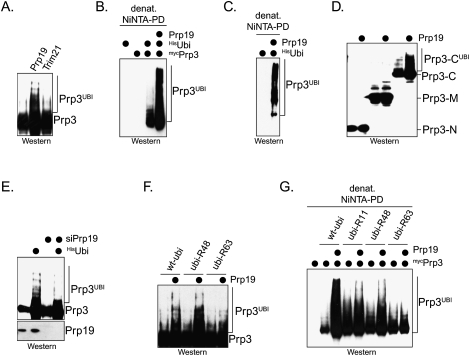
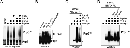
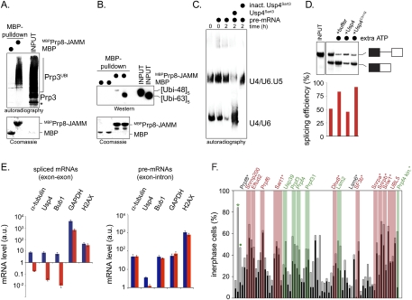
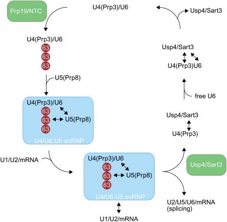
The spliceosome, a dynamic assembly of proteins and RNAs, catalyzes the excision of intron sequences from nascent mRNAs. Recent work has suggested that the activity and composition of the spliceosome are regulated by ubiquitination, but the underlying mechanisms have not been elucidated. Here, we report that the spliceosomal Prp19 complex modifies Prp3, a component of the U4 snRNP, with nonproteolytic K63-linked ubiquitin chains. The K63-linked chains increase the affinity of Prp3 for the U5 snRNP component Prp8, thereby allowing for the stabilization of the U4/U6.U5 snRNP. Prp3 is deubiquitinated by Usp4 and its substrate targeting factor, the U4/U6 recycling protein Sart3, which likely facilitates ejection of U4 proteins from the spliceosome during maturation of its active site. Loss of Usp4 in cells interferes with the accumulation of correctly spliced mRNAs, including those for alpha-tubulin and Bub1, and impairs cell cycle progression. We propose that the reversible ubiquitination of spliceosomal proteins, such as Prp3, guides rearrangements in the composition of the spliceosome at distinct steps of the splicing reaction.


Figures

Figure 1.
Figure 1.
Usp4 is required for faithful cell cycle progression. (A) Identification of human DUBs that regulate the cell cycle. Candidate DUBs identified in three parallel siRNA screens were depleted in HeLa cells by four independent siRNAs. HeLa cells were treated with taxol, and, 24 h later, the percentage of cells arrested prior to mitosis (black bar) and the number of cells unable to maintain a spindle checkpoint arrest (gray bar) were determined. siRNA against luciferase (*) and against Usp44 (•) were used as negative and positive controls, respectively. A percentage of >30% nonmitotic cells was set as an arbitrary threshold for specificity (red line). DUBs whose depletion by at least two siRNAs predominantly results in premitotic arrest are labeled in red, whereas DUBs required for a stable checkpoint response are labeled in green. (B) The efficiency of Usp4 depletion matches the strength of its cell cycle phenotypes. Five different siRNAs against Usp4 were tested for effects on cell cycle progression (left panel) and efficiency of mRNA depletion (right panel), as determined by RT–PCR. The abundance of mRNAs is shown in comparison with untransfected (utf) control cells. (C) siRNAs against the 3′-untranslated region (UTR) of Usp4 deplete the Usp4 protein from cells. Two independent siRNAs against the 3′-UTR of Usp4 were tested for their effect on depletion of Usp4, compared with the loading control β-actin, as determined by Western blot. (D) The activity of Usp4 as a DUB is required for its role in cell cycle control. HeLa cells were treated against siRNA against the 3′-UTR of Usp4, and the number of mitotic cells with errors in chromosome segregation (black bars) or spindle formation (gray bars) was determined. When indicated, cells were also transfected with vectors encoding siRNA-resistant Usp4 or the catalytically inactive Usp4C311A. (E) Usp4 does not directly counteract the E3s APC/C or Trim21. HeLa treated with siRNA against Usp4 and taxol were scored for the percentage of interphase cells. When indicated, siRNAs against UbcH10, Ube2S, or Trim21 were cotransfected.
Figure 2.
Figure 2.
Sart3 is a targeting factor of Usp4. (A) Identification of Sart3 as a binding partner of Usp4. The regulatory domain of Usp4 was fused to MBP (MBPUsp4-NT; amino acids 1–296). Immobilized MBP and MBPUsp4-NT were incubated with extracts of mitotic HeLa S3 cells (CP extracts). Proteins retained by MBPUsp4-NT, but not MBP, were detected by Coomassie staining and identified by mass spectrometry. The asterisks mark proteins that were retained by both MBP and MBPUsp4-NT; we did not identify these unspecific binding partners. (B) Schematic overview of Sart3 and its interaction with Usp4. Sart3 consists of seven HAT repeats, one coiled-coil, two RNA recognition motifs, and one LSM domain. The HAT repeat 7 of Sart3 is required for the interaction with the partially overlapping DUSP and DUF1055 domains of Usp4. (C) Sart3 binds Usp4 in vivo. HeLa cells were transfected with mycSart3 and HAUsp4, and were grown asynchronously or arrested in mitosis by nocodazole. HAUsp4 was purified on αHA-agarose, and coprecipitating mycSart3 was detected by αmyc-Western blot. (D) Sart3 binds endogenous Usp4. HASart3 was expressed in HeLa cells and purified on αHA-agarose. Coprecipitating endogenous Usp4 was detected by Western blot using αUsp4 antibodies. (E) Usp4 and Sart3 interact under physiological conditions. We generated a stable U2OS cell line that inducibly expresses FlagSart3 at low concentrations upon addition of doxycycline to the medium. FlagSart3 was purified by αFlag immunoprecipitation, and coprecipitating endogenous Usp4 was detected by Western blotting using αUsp4 antibodies. The asterisk denotes a nonspecific band. (F) The HAT7 domain of Sart3 is required for the interaction with Usp4. HeLa cells were cotransfected with HA-tagged deletion mutants of Sart3 and mycUsp4, and the interaction of the Sart3 mutants with Usp4 was analyzed by αHA-affinity purification. Coprecipitating mycUsp4 was detected by αmyc-Western blotting. Expression of Sart3ΔHAT4–6 appeared to be toxic in HeLa cells, complicating the interpretation of results with this mutant. (G) The interaction with Usp4 is required for the role of Sart3 as a cell cycle regulator. Sart3 was depleted from HeLa cells using siRNA against the 3′-UTR of Sart3, and the percentage of mitotic cells with obvious chromosome missegregation or spindle defects was determined. As indicated, cells were cotransfected with siRNA-resistant Sart3 or deletion mutants. Similar to Usp4, depletion of Sart3 results in chromosome missegregation (black bars). The Usp4-binding-deficient Sart3ΔHAT7 is unable to rescue these phenotypes. (H) Sart3 does not activate Usp4. The DUB activity of Usp4 was measured by monitoring the release of the fluorophore AMC from the C terminus of ubiquitin, which results in an increase of the fluorescence signal at 469 nm. The activities of Usp4, Usp4 inactivated with NEM, and the Usp4Sart3 complex were compared. (I) Sart3 functions as a targeting factor of Usp4. The intracellular localization of mycUsp4 was analyzed by fluorescence microscopy. Expression of Sart3 (as indicated on the left) results in nuclear translocation of Usp4. Coexpression of the Usp4-binding-deficient Sart3ΔHAT7 or the nuclear protein Prp3 had no effect on the localization of Usp4.
Figure 3.
Figure 3.
Sart3 recruits Prp3 to Usp4. (A) Depletion of Prp3 causes a cell cycle defect. HeLa cells were transfected with siRNA against Mad2, Usp4, Sart3, and Prp3 and treated with taxol. After 24 h, the percentage of cells in mitosis (black bars) and interphase (gray bars) was determined by microscopy. (B) Depletion of Prp3 results in chromosome missegregation. HeLa cells were transfected with siRNA against Prp3 or Usp4. As indicated, siRNA-resistant cDNA encoding Prp3 or Usp4 was cotransfected. Mitotic cells were analyzed for chromosome missegregation and spindle defects after immunofluorescence against α-tubulin and DNA. (C) Sart3 recruits Prp3 to Usp4. MBP or MBPUsp4 was immobilized on amylose resin and incubated with 35S-Sart3 or 35S-Prp3. As indicated, recombinant HisPrp3 or HisSart3 was added. Bound proteins were detected by autoradiography. Radiolabeled Prp3 interacts with MBPUsp4 only in the presence of HisSart3. (D) Sart3 recruits Usp4 to Prp3. MBP or MBPPrp3 was immobilized on amylose resin and incubated with 35S-Usp4 or 35S-Sart3. When indicated, recombinant HisSart3 or HisUsp4 was added, and bound proteins were detected by autoradiography. HisSart3 comigrates with MBPPrp3. The asterisk denotes an unknown protein in reticulocyte lysate that interacts with MBPPrp3. (E) The N terminus of Sart3 binds Prp3. HASart3 or the indicated deletion mutants were coexpressed with mycPrp3, and were affinity-purified on αHA-agarose. Bound mycPrp3 was detected by αmyc-Western blotting. Note that Sart3286-705, which efficiently interacts with Usp4, fails to bind Prp3. (F) Coexpression of Sart3 increases the efficiency of the Usp4–Prp3 interaction. HeLa cells were cotransfected with HAUsp4, mycPrp3, and, as indicated, Sart3. Usp4 complexes were affinity-purified on αHA-agarose, and bound mycPrp3 was detected by αmyc-Western blot. (G) Sart3 triggers the colocalization of Prp3 and Usp4. The localization of HAUsp4 (red) and mycPrp3 (green) in HeLa cells was analyzed by fluorescence microscopy in the absence (top panels) or presence (bottom panels) of coexpressed Sart3.
Figure 4.
Figure 4.
Prp3 is ubiquitinated by the Prp19 complex in vitro. (A) Prp19 associates with Prp3 in vitro. MBP, MBPPrp3, and MBP-tagged truncation mutants of Prp3 were immobilized on amylose resin, and were incubated with 35S-Prp19. Bound Prp19 was detected by autoradiography. Purified MBPPrp3-M does not bind efficiently to beads, and thus cannot be analyzed (its position is marked by an asterisk). (B) Prp19 associates with Prp3 in vivo. HeLa cells were cotransfected with HAPrp3 and mycPrp19. HAPrp3 was affinity-purified on αHA-agarose, and copurifying Prp19 was detected by αmyc-Western blot. When indicated, cells were synchronized in mitosis with nocodazole prior to the immunoprecipitation. (C) The Prp19 complex ubiquitinates Prp3 in vitro. The Prp19 complex was affinity-purified from HeLa S3 cells by using αPrp19-agarose or αCdc5-agarose. Complexes were analyzed by silver staining (left panel) or Western blotting (bottom left panel) using specific antibodies against Prp19 and Prp3. Beads were incubated with 35S-Prp3 for 2 h. Unbound proteins were washed away, before the NTC/Prp3 complexes were incubated with purified E1, UbcH5c, ubiquitin, and energy mix. Ubiquitinated Prp3 was detected by autoradiography. The asterisk marks an N-terminally truncated Prp3 resulting from alternative start codon usage in the IVT/T. (D) The Prp19 complex forms ubiquitin chains on Prp3. 35S-Prp3 was ubiquitinated by the Prp19 complex and UbcH5c in the presence of ubiquitin or methylubiquitin, which is unable to support ubiquitin chain formation. Ubiquitinated Prp3 was detected by autoradiography. The asterisk marks an N-terminally truncated Prp3 resulting from alternative start codon usage in the IVT/T reaction. (E) The Prp19 complex can assemble K63-linked ubiquitin chains in vitro. The Prp19 complex was used to catalyze the ubiquitination of 35S-Prp3 in the presence of ubiquitin or ubiquitin with Lys63 as its only lysine residue (ubi-K63). As E2s, UbcH5c, Ube2N/UEV1A, or UbcH5c and Ube2N/UEV1A were used. Ubiquitinated Prp3 was detected by autoradiography. The asterisk marks an N-terminally truncated Prp3 resulting from alternative start codon usage in the IVT/T reaction. (F) The Prp19 complex requires the hydrophobic patch on ubiquitin to support chain formation on Prp3. Ubiquitination of 35S-Prp3 was catalyzed by the Prp19 complex in the presence of ubiquitin or the mutant ubi-I44A. As a comparison, the ubiquitination of Prp3 was also performed in the presence of its DUB, Usp4Sart3. Ubiquitinated Prp3 was detected by autoradiography. The asterisk marks an N-terminally truncated Prp3 resulting from alternative start codon usage in the IVT/T reaction. (G) The Prp19 complex ubiquitinates the C-terminal domain of Prp3. 35S-Prp3 or truncation mutants (Prp3-N, Prp3-M, or Prp3-C) were tested for ubiquitination by affinity-purified Prp19 and UbcH5c. Ubiquitinated Prp3 was detected by autoradiography. Note that only Prp3-C, but neither Prp3-N nor Prp3-M, is ubiquitinated in a Prp19-dependent manner. The asterisk marks a truncation product of the Prp3-C construct.
Figure 5.
Figure 5.
The Prp19 complex (NTC) promotes the ubiquitination of Prp3 in vivo. (A) Expression of Prp19 triggers the modification on Prp3 in cells. mycPrp3 was coexpressed with Prp19 or the E3 Trim21 in HeLa cells, and was analyzed for modifications by αmyc-Western blot. (B) Prp19 promotes the ubiquitination of Prp3 in HeLa cells. mycPrp3, Prp19, and Hisubiquitin were expressed in HeLa cells as indicated. HisUbiquitin and covalently modified proteins were purified from cells under denaturing conditions on NiNTA-agarose. Copurifying ubiquitinated mycPrp3 was detected by αmyc-Western blot. (C) The NTC promotes the ubiquitination of endogenous Prp3 in cells. Prp19 and Hisubiquitin were expressed in HeLa cells as indicated, and ubiquitinated proteins were purified on NiNTA-agarose under denaturing conditions. Ubiquitinated endogenous Prp3 was detected by Western blotting using specific αPrp3 antibodies. (D) The NTC ubiquitinates the C-terminal domain of Prp3 in cells. The myc-tagged truncation mutants Prp3-N, Prp3-M, and Prp3-C were expressed in HeLa cells. Where indicated, Prp19 was coexpressed, and the modification of the Prp3 proteins was analyzed by αmyc-Western blot. (E) Depletion of Prp19 by siRNA reduces the ubiquitination of Prp3 in HeLa cells. The ubiquitination of mycPrp3 was analyzed in HeLa cells by αmyc-Western blot, after Prp19 was depleted by a specific siRNA targeting its 3′-UTR. (F) The NTC promotes the modification of Prp3 with K63-linked chains in cells. mycPrp3 and Prp19 were expressed in HeLa cells, as indicated. The coexpression was performed in the presence of wt-ubi, ubi-R48 (which has Lys48 mutated to Arg), or ubi-R63. (G) The NTC promotes the modification of Prp3 with K63-linked chains, as detected by denaturing purification of ubiquitin conjugates. mycPrp3 and Prp19 were expressed in HeLa cells with the indicated Hisubiquitin mutants. Ubiquitin conjugates were purified by denaturing NiNTA pull-down, and mycPrp3 was detected by αmyc-Western blotting.
Figure 6.
Figure 6.
Usp4Sart3 counteracts the NTC. (A) Usp4Sart3 deubiquitinates Prp3 in vitro. 35S-Prp3 was ubiquitinated by affinity-purified NTC (Prp19), and was subsequently incubated with Usp4 or Usp4Sart3. Control reactions with NEM-inactivated Usp4 or Usp4Sart3 were performed in parallel. The ubiquitination of Prp3 was analyzed by autoradiography. The asterisk marks an N-terminally truncated Prp3 resulting from alternative start codon usage in the IVT/T reaction. (B) Usp4 counteracts Prp19 in vivo. mycPrp3 was expressed with Prp19, Sart3, and Usp4Sart3, as indicated, and was analyzed for modifications by αmyc-Western blot. (C) Usp4 counteracts the NTC in vivo, as detected by denaturing purification of ubiquitinated proteins. mycPrp3 was expressed with Hisubiquitin, Prp19, and Usp4Sart3, and covalently modified proteins were purified under denaturing conditions on NiNTA-agarose. Ubiquitinated mycPrp3 was detected by αmyc-Western blot. (D) Deubiquitination of Prp3 requires the catalytic activity of Usp4. mycPrp3 was coexpressed with Hisubiquitin, Prp19, and either Usp4Sart3, Usp4C311A/Sart3, or Usp44. Covalently modified proteins were purified on NiNTA-agarose under denaturing conditions, and ubiquitinated mycPrp3 was detected by αmyc-Western blotting.
Figure 7.
Figure 7.
Ubiquitin-dependent regulation of the spliceosome. (A) The U5 component Prp8 recognizes ubiquitinated Prp3. 35S-Prp3 was ubiquitinated by the NTC and UbcH5c, and was incubated with immobilized MBP and MBPPrp8-JAMM (the isolated JAMM domain). Bound proteins were detected by autoradiography. The asterisk marks an N-terminally truncated Prp3 resulting from alternative start codon usage in the IVT/T reaction. (B) Prp8 preferentially interacts with K63-linked chains. MBP and MBPPrp8-JAMM were coupled to amylose resin and incubated with K48- or K63-linked ubiquitin pentamers, as indicated. Bound ubiquitin proteins were visualized by αubiquitin-Western blot. (C) Usp4Sart3 regulates the stability of the U4/U6.U5 snRNP in HeLa splicing extracts. HeLa splicing extracts were supplemented with Ftz-pre mRNA, Usp4Sart3, or NEM-treated, inactive Usp4Sart3, as indicated. The abundance of the U4/U6.U5 and U4/U6 snRNPs was monitored by Northern blotting using U6 snRNA as a probe (Konarska and Sharp 1987). (D) Usp4Sart3 regulates splicing in vitro. HeLa splicing extracts were prepared in the presence of ATP to maintain a functional ubiquitin system (Williamson et al. 2009). The splicing of radiolabeled Ftz-pre-mRNA was monitored by autoradiography in the presence of additional ATP, Usp4Sart3, or catalytically inactive Usp4C311S/Sart3, and gel bands were quantitated using ImageQuant software. The percent splicing efficiency was calculated as spliced mRNA over total mRNA (pre-mRNA + spliced mRNA). (E) Usp4 is required for splicing in cells. HeLa cells were treated with siRNA against Usp4, and were arrested in mitosis with nocodazole. The abundance of mature mRNAs against the indicated targets was determined by qPCR. (Blue bars) Control cells; (red bars) Usp4-depleted cells; (a.u.) arbitrary units derived from the cT-value. Primers annealed to either exon junction sequences or the single exon of H2AX. In a control qPCR experiment, primers annealed to the one of the gene's exons and neighboring introns. (F) Loss of several spliceosomal proteins results in cell cycle defects also observed upon Usp4 depletion. Spliceosomal proteins were depleted from HeLa cells by siRNA. HeLa cells were treated with taxol, and the percentage of cells showing premitotic arrest or spindle checkpoint bypass was determined. Positive hits are indicated above, and those marked with an asterisk were identified previously in siRNA screens. Depletions resulting predominantly in premitotic arrest are labeled in red, and those with a significant spindle checkpoint bypass phenotype are marked in green.
Figure 8.
Figure 8.
Model of the ubiquitin-dependent regulation of the U4/U6.U5 snRNP. The Prp19 complex (NTC) promotes the modification of Prp3, a U4 component, with K63-linked ubiquitin chains (red circles). Ubiquitinated Prp3 can be recognized by the JAMM domain of Prp8, a U5 component, thereby allowing for the stabilization of the U4/U6.U5 snRNP. After docking of the U4/U6.U5 snRNP at the spliceosome, U4 snRNA and the U4 proteins dissociate along with U1 snRNA and proteins of the U6 snRNP. We propose that deubiquitination of Prp3 by Usp4Sart3 decreases its affinity to Prp8 and facilitates dissociation. Sart3 also promotes the reannealing of U4 and U6 snRNPs, allowing the U4/U6 snRNP to enter another round of splicing.

Similar articles

Cited by

References

    1. Aravind L, Koonin EV 2000. The U box is a modified RING finger—A common domain in ubiquitination. Curr Biol 10: R132–R134 doi: 10.1016/S0960-9822(00)00398-5 - PubMed
    1. Bell M, Schreiner S, Damianov A, Reddy R, Bindereif A 2002. p110, a novel human U6 snRNP protein and U4/U6 snRNP recycling factor. EMBO J 21: 2724–2735 - PMC - PubMed
    1. Bellare P, Kutach AK, Rines AK, Guthrie C, Sontheimer EJ 2006. Ubiquitin binding by a variant Jab1/MPN domain in the essential pre-mRNA splicing factor Prp8p. RNA 12: 292–302 - PMC - PubMed
    1. Bellare P, Small EC, Huang X, Wohlschlegel JA, Staley JP, Sontheimer EJ 2008. A role for ubiquitin in the spliceosome assembly pathway. Nat Struct Mol Biol 15: 444–451 - PMC - PubMed
    1. Burns CG, Ohi R, Mehta S, O'Toole ET, Winey M, Clark TA, Sugnet CW, Ares M Jr, Gould KL 2002. Removal of a single α-tubulin gene intron suppresses cell cycle arrest phenotypes of splicing factor mutations in Saccharomyces cerevisiae. Mol Cell Biol 22: 801–815 - PMC - PubMed

Publication types

MeSH terms