Review
. 2011 Apr;32(4):617-26.
doi: 10.3174/ajnr.A2144. Epub 2010 Jul 1.

Sphenoid masses in children: radiologic differential diagnosis with pathologic correlation

Affiliations
Review

Sphenoid masses in children: radiologic differential diagnosis with pathologic correlation

Y W Lui et al. AJNR Am J Neuroradiol. 2011 Apr.

Abstract

Childhood central skull base masses are rare, often difficult to diagnose, and have overlapping imaging findings. In this review, we provide an overview of the epidemiology, clinical findings, and management of pediatric sphenoid bone and sphenoid sinus masses with an emphasis on imaging findings that may help to differentiate lesions. Radiologic-pathologic correlation is provided. Finally, an imaging-based algorithm is presented as a guide to help radiologists narrow their differential diagnoses. Some of the entities discussed are virtually unique to the pediatric population; others occur rarely in this age group but should be considered in the appropriate clinical setting. Entities included in the discussion are grouped into 2 categories: those that cause nonaggressive osseous remodeling and those that are more commonly associated with aggressive bone changes. Mucocele, aneurysmal bone cyst, giant cell lesions, meningioma, and fibrous dysplasia tend to remodel bone, while entities such as chordoma, craniopharyngioma, rhabdomyosarcoma, sinonasal carcinoma, and neuroblastoma may cause more aggressive local bone changes.


Figures

Fig 1.
Fig 1.
An imaging-guided algorithm.
Fig 2.
Fig 2.
An 18-year-old man with sphenoid mucocele. A, Axial T2WI demonstrates a fluid-filled and expanded sphenoid sinus. B, Sagittal noncontrast T1WI shows a fluid/debris level within the lesion. Relative hyperintense material on T1WI reflects proteinaceous fluid content. There is intracranial extension with loss of the cortical margin of the planum sphenoidale (arrow).
Fig 3.
Fig 3.
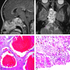
A 19-year-old woman with aneurysmal bone cyst. A and B, Gadolinium-enhanced sagittal T1WI (A) and axial T2WI (B) show multiple fluid/fluid levels in a complex multicystic mass centered on the sphenoid bone, expanding the bone and filling the sphenoid sinus. C, Photomicrograph shows blood-filled cavernous channels at low-power magnification with (HE staining, original magnification ×4). D, Higher power photomicrograph shows spindle cells in the vessel walls, reactive bone formation, and multinucleated giant cells typical of these lesions (HE staining, original magnification ×40).
Fig 4.
Fig 4.
Two patients with giant cell lesions of the sphenoid bone. A, An 18-year-old man with sphenoid giant cell tumor: axial T2WI shows relative hypointense areas within a lesion centered on the sphenoid bone and obliterating the sphenoid sinus cavity. These hypointense areas may represent areas of old blood products and fibrotic change. The carotids are displaced laterally (arrows). B, Gadolinium-enhanced sagittal T1WI shows the lesion avidly enhancing (black arrow). The pituitary gland and sella are displaced cephalad (white arrow). C, A 27-year-old woman with giant cell reparative granuloma. Coronal reformatted image in a soft-tissue window from a noncontrast CT scan shows a soft-tissue mass centered on the sphenoid sinus with dehiscence of the planum sphenoidale (arrow). D, HE stain on the photomicrograph shows giant cells with multiple nuclei, fibrous tissue, and blood products characteristic of this reactive lesion (original magnification ×40).
Fig 5.
Fig 5.
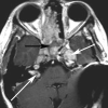
A 10-year-old female patient with central skull base meningioma invading the paranasal sinuses. Axial postcontrast T1WI demonstrates an enhancing mass within the central skull base. The mass involves the ethmoid and sphenoid sinuses and was a biopsy-proved meningioma. Nonenhancing anterior portion of the sphenoid sinus is filled with secretions (black arrow). Multiple additional schwannomas are seen in the Meckel cave and the internal auditory canal (white arrows) in this patient with neurofibromatosis type 2. Although meningioma is common in adults, in pediatric patients, the lesion is much rarer and occurs in the setting of neurofibromatosis type 2 or prior radiation therapy.
Fig 6.
Fig 6.
A 13-year-old girl with fibrous dysplasia affecting the sphenoid bone. A, Bone window on CT shows classic ground-glass attenuation in the sphenoid bone. B, The sphenoid sinuses are small and demonstrate mucosal disease. The lesion is hypointense on T2WI. C, This MR imaging appearance is likely attributable to the fibrous stroma, illustrated on the photomicrograph with HE stain demonstrating woven bone embedded in a spindle cell stroma (original magnification ×4). Fibrous dysplasia can have extreme heterogeneity on MR imaging and can be a confusing lesion to diagnose. Correlation with CT should always be done.
Fig 7.
Fig 7.
A 4-year-old girl with hypothalamic astrocytoma. A, Sagittal postcontrast T1WI shows a large enhancing mass extending from the suprasellar region. The lesion expands and fills the sella with remodeling of the sphenoid bone. The sphenoid sinus is small (black arrow). Postoperative changes and artifacts are present (white arrow). B and C, Compact and spongy areas are seen on photomicrographs of histopathologic specimens, correlating to solid and cystic components of the tumor (HE staining; original magnification ×40).
Fig 8.
Fig 8.
Two patients with chordoma involving the sphenoid bone. A, An 18-year-old male patient. Axial T2WI shows a multicystic lesion involving the central skull base, including the sphenoid bone, sphenoid sinus, posterior ethmoids, and left middle cranial fossa. Focal areas of T2 hyperintensity are seen. B, In the coronal reformatted image, a bone window shows dehiscence of the planum sphenoidale (arrow). C, An 11-year-old boy with chordoma. Sagittal noncontrast T1WI shows the lesion to nearly fill the sphenoid sinus. There is a small residual aerated portion of the sphenoid sinus cavity (arrow). D, Photomicrograph shows that neoplastic epithelioid cells form cords in a mucoid background (HE staining; original magnification ×40).
Fig 9.
Fig 9.
A 12-year-old boy with craniopharyngioma. A, Axial fluid-attenuated inversion recovery image shows the cystic portion of the tumor (black arrow) within the central sphenoid bone (white arrow). There is a fluid/fluid level. B, Photomicrograph of a histopathologic specimen shows findings characteristic of craniopharyngioma, with nests of anucleated squamous cells embedded in fibrous tissue and clefts consistent with cholesterol (HE staining; original magnification ×10).
Fig 10.
Fig 10.
A 16-year-old boy with rhabdomyosarcoma. A, Bone window from a noncontrast CT examination show opacification of the right-sided paranasal sinuses, including the right sphenoid sinus (arrow). There are areas of dehiscent bone involving the maxillary sinus walls. B, Soft-tissue window shows the lesion involving the right nasal cavity, right maxillary and sphenoid sinuses, right orbit, and right pterygopalatine fossa (white arrow). Note the normal fat within the left pterygopalatine fossa (black arrow).
Fig 11.
Fig 11.
A 16-year-old boy with juvenile angiofibroma. A, Axial unenhanced CT scan demonstrates widening of the right sphenopalatine foramen with abnormal soft tissue obliterating the normal fat within the pterygopalatine fossa (white arrow). Soft tissue extends anteriorly into the posterior nasopharynx and posteriorly into the sphenoid bone and right sphenoid sinus (black arrow). B, Lateral projection from a right external carotid artery angiogram in the arterial phase demonstrates an attenuated tumor blush and an enlarged internal maxillary artery with tortuous vessels in the right pterygopalatine fossa (arrow).
Fig 12.
Fig 12.
A 20-year-old man with sinonasal carcinoma. A, Bone window from an unenhanced CT scan shows destructive osseous changes (arrow). B, Soft tissue window at the same level shows an associated soft tissue mass in the right central skull base.
Fig 13.
Fig 13.
A 3-year-old girl with neuroblastoma. A, Sagittal postcontrast T1WI shows a mass centered on the sphenoid bone, an unusual location for a neuroblastoma metastasis, obliterating the sphenoid sinus. There is mass effect on the gyrus rectus. B, A multilobulated mass demonstrates characteristic relative hypointense signal intensity on an axial T2WI. The lesion has an intracranial component, extending along the anterior aspect of the middle cranial fossae bilaterally (arrows). C, The left adrenal primary is shown on coronal postcontrast T1WI of the abdomen.

Similar articles

Cited by

References

    1. Levine H. The sphenoid sinus, the neglected nasal sinus. Arch Otolaryngol 1978;104:585–87 - PubMed
    1. Grossman RI, Yousem DM. Neuroradiology: The Requisites. St. Louis:Mosby-Year Book Inc;1994
    1. Alvarez RJ, Liu NJ, Isaacson G. Pediatric ethmoid mucoceles in cystic fibrosis: long-term follow-up of reported cases. Ear Nose Throat J 1997;76:538–39,543–46 - PubMed
    1. Bailey BJ, Johnson JT, Newlands SD, eds. Head & Neck Surgery: Otolaryngology. 4th ed. Philadelphia:Lippincott Williams & Wilkins;2006
    1. Stankiewicz JA. Sphenoid sinus mucocele. Arch Otolaryngol Head Neck Surg 1989;115:735–40 - PubMed