Review
. 2010 Jul;2(7):a001180.
doi: 10.1101/cshperspect.a001180. Epub 2010 Apr 7.

Pathologies associated with the p53 response

Affiliations
Review

Pathologies associated with the p53 response

Andrei V Gudkov et al. Cold Spring Harb Perspect Biol. 2010 Jul.

Abstract

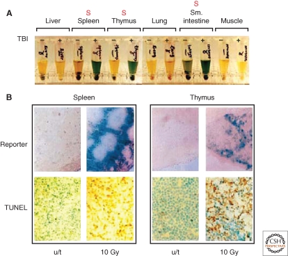
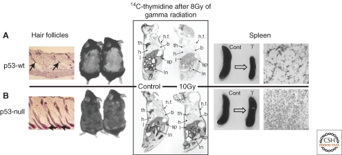
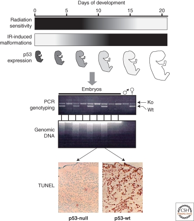
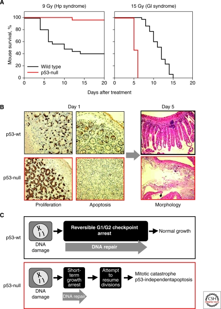
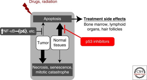
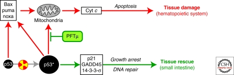
Although p53 is a major cancer preventive factor, under certain extreme stress conditions it may induce severe pathologies. Analyses of animal models indicate that p53 is largely responsible for the toxicity of ionizing radiation or DNA damaging drugs contributing to hematopoietic component of acute radiation syndrome and largely determining severe adverse effects of cancer treatment. p53-mediated damage is strictly tissue specific and occurs in tissues prone to p53-dependent apoptosis (e.g., hematopoietic system and hair follicles); on the contrary, p53 can serve as a survival factor in tissues that respond to p53 activation by cell cycle arrest (e.g., endothelium of small intestine). There are multiple experimental indications that p53 contributes to pathogenicity of acute ischemic diseases. Temporary reversible suppression of p53 by small molecules can be an effective and safe approach to reduce severity of p53-associated pathologies.


Figures

Figure 1.
Figure 1.
Tissue specificity of p53 activity. (A) Detection of β-galactosidase reporter activity in indicated tissue extracts from transgenic mice carrying p53-responsive lacZ and treated with 10 Gy of total body irradiation. Reporter activity is seen only in radiosensitive organs (marked with red “S”). (B) Massive apoptosis in tissues demonstrating strong response of p53-dependent reporter 8 h after irradiation detected using TUNEL staining for DNA fragmentation in situ. For details, see (Komarova et al. 1997).
Figure 2.
Figure 2.
Biological effects driven by p53 in vivo. Left panel: mouse model of chemotherapy-induced alopecia shows resistance of hair follicles of p53-null mice to cyclophosphamide-induced apoptosis accompanied with lack of hair loss (Botchkarev et al. 2001). Middle panel: DNA replication block observed shortly after TBI (figure shows results obtained 24 h postirradiation) is p53-specific and is not seen in p53-null mice (Komarova et al. 2000). Right panel: massive cell loss occurring in the spleen 24 h post TBI (example shown for 10 Gy) because of massive apoptosis is p53-specific and is undetectable in p53-null mice (Komarova et al. 1997).
Figure 3.
Figure 3.
Radiosensitivity of early embryos is p53-driven (Komarova et al. 1997). Upper panel: schematic description of the correlation among the time stage-dependence of resistance to TBI (black-higher sensitivity), frequency of radiation-induced malformations and reduction of p53 occurring in mid gestation. Lower panel: differential response of embryos to TBI of pregnant female depending on their p53 status. Embryos were either p53-null or p53+/− because they originated from p53-null female and p53+/− male as shown by PCR genotyping. Massive apoptosis detected by TUNEL assay and by electrophoretic assessment of DNA degradation was restricted to those embryos that possess wild-type p53 allele.
Figure 4.
Figure 4.
p53 is a survival factor in irradiated small intestine (Komarova et al. 2004). (A) p53-null mice are resistant to doses of TBI that induce hematopoietic (HP) but are hypersensitive to gastrointestinal (GI) acute radiation syndrome (ARS). (B) Complete destruction of the epithelium of small intestine in p53-null mice by day 5 after 15 Gy of TBI follows continuous proliferation and lack of early apoptosis observed on day 1 (8 h) post TBI. Growth arrest (determined by BrdU incorporation) and massive apoptosis in the crypts of p53 wild-type mice following TBI are associated with better preserved organ structure 5 d later. (C) Schematic interpretation of the earlier described results, which attributes radioresistance of p53 wild type small intestine to growth arrest function of p53.
Figure 5.
Figure 5.
Schematic description of the strategy of pharmacological inhibition of p53 to protect mammalian organism from tissue specific toxicities associated with systemic genotoxic stresses.
Figure 6.
Figure 6.
Existence of two branches in the p53 pathway allows pharmacological dissection of different p53 activities. By selective blocking of the mitochondrial branch of the pathway (by PFTμ) can protect from lethal hematopoietic radiation syndrome without affecting transactivation-mediated functions of p53 (growth arrest at cell cycle checkpoints, DNA repair, etc.), thus preserving tissue protective functions of p53. For details, see Strom et al. (2006).

References

    1. Akhtar RS, Geng Y, Klocke BJ, Roth KA 2006. Neural precursor cells possess multiple p53-dependent apoptotic pathways. Cell Death Differentiation 13:1727–1739 - PubMed
    1. Alvarez-Salas LM, Arpawong TE, DiPaolo JA 1999. Growth inhibition of cervical tumor cells by antisense oligodeoxynucleotides directed to the human papillomavirus type 16 E6 gene. Antisense Nucleic Acid Drug Dev 9:441–450 - PubMed
    1. Alves da Costa C, Mattson MP, Ancolio K, Checler F 2003. The C-terminal fragment of presenilin 2 triggers p53-mediated staurosporine-induced apoptosis, a function independent of the presenilinase-derived N-terminal counterpart. J Biol Chem 278:12064–12069 - PubMed
    1. Armstrong JF, Kaufman MH, Harrison DJ, Clarke AR 1995. High-frequency developmental abnormalities in p53-deficient mice. CurrBiol 5:931–936 - PubMed
    1. Aylon Y, Oren M 2007. Living with p53, dying of p53. Cell 130:597–600 - PubMed

Publication types

Substances