

Basal ganglia surface morphology and the effects of stimulant medications in youth with attention deficit hyperactivity disorder
- PMID: 20595414
- PMCID: PMC4254769
- DOI: 10.1176/appi.ajp.2010.09091259
Basal ganglia surface morphology and the effects of stimulant medications in youth with attention deficit hyperactivity disorder
Abstract
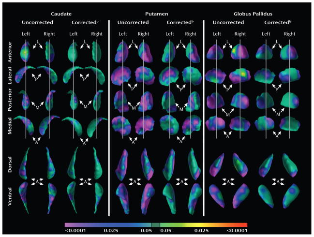
Objective: Disturbances in the basal ganglia portions of cortico-striato-thalamo-cortical circuits likely contribute to the symptoms of attention deficit hyperactivity disorder (ADHD). The authors examined the morphologic features of the basal ganglia nuclei (caudate, putamen, and globus pallidus) in children with ADHD.
Method: A total of 104 individuals (combined-type ADHD patients: N=47; healthy comparison subjects: N=57), aged 7 to 18 years, were examined in a cross-sectional case-control study using anatomical magnetic resonance imaging. Conventional volumes and the surface morphology for the basal ganglia were measured.
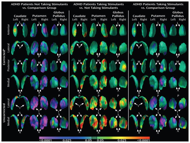
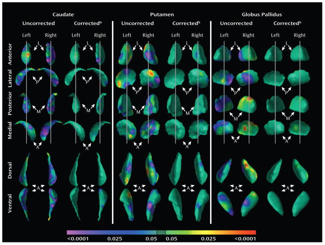
Results: Overall volumes were significantly smaller only in the putamen. Analysis of the morphological surfaces revealed significant inward deformations in each of the three nuclei, localized primarily in portions of these nuclei that are components of limbic, associative, and sensorimotor pathways in the cortico-striato-thalamo-cortical circuits in which these nuclei reside. The more prominent these inward deformations were in the patient group, the more severe the ADHD symptoms. Surface analyses also demonstrated significant outward deformations of all basal ganglia nuclei in the ADHD children treated with stimulants compared with those ADHD youth who were untreated. These stimulant-associated enlargements were in locations similar to the reduced volumes detected in the ADHD group relative to the comparison group. The outward deformations associated with stimulant medications attenuated the statistical effects of the primary group comparisons.
Conclusions: These findings potentially represent evidence of anatomical dysregulation in the circuitry of the basal ganglia in children with ADHD and suggest that stimulants may normalize morphological features of the basal ganglia in children with the disorder.
Figures
Similar articles
-
Basal ganglia volume and shape in children with attention deficit hyperactivity disorder.Am J Psychiatry. 2009 Jan;166(1):74-82. doi: 10.1176/appi.ajp.2008.08030426. Epub 2008 Nov 17. Am J Psychiatry. 2009. PMID: 19015232 Free PMC article.
-
Asymmetry of basal ganglia in children with attention deficit hyperactivity disorder.Neuro Endocrinol Lett. 2007 Oct;28(5):604-9. Neuro Endocrinol Lett. 2007. PMID: 17994006
-
Developmentally stable whole-brain volume reductions and developmentally sensitive caudate and putamen volume alterations in those with attention-deficit/hyperactivity disorder and their unaffected siblings.JAMA Psychiatry. 2015 May;72(5):490-9. doi: 10.1001/jamapsychiatry.2014.3162. JAMA Psychiatry. 2015. PMID: 25785435
-
Meta-analysis of structural MRI studies in children and adults with attention deficit hyperactivity disorder indicates treatment effects.Acta Psychiatr Scand. 2012 Feb;125(2):114-26. doi: 10.1111/j.1600-0447.2011.01786.x. Epub 2011 Nov 28. Acta Psychiatr Scand. 2012. PMID: 22118249 Review.
-
[Structural and functional neuroanatomy of attention-deficit hyperactivity disorder (ADHD)].Encephale. 2009 Apr;35(2):107-14. doi: 10.1016/j.encep.2008.01.005. Epub 2008 Jul 7. Encephale. 2009. PMID: 19393378 Review. French.
Cited by
-
Reduced basal ganglia tissue-iron concentration in school-age children with attention-deficit/hyperactivity disorder is localized to limbic circuitry.Exp Brain Res. 2022 Dec;240(12):3271-3288. doi: 10.1007/s00221-022-06484-7. Epub 2022 Oct 27. Exp Brain Res. 2022. PMID: 36301336 Free PMC article.
-
Gray matter alterations in adults with attention-deficit/hyperactivity disorder identified by voxel based morphometry.Biol Psychiatry. 2011 May 1;69(9):857-66. doi: 10.1016/j.biopsych.2010.09.053. Epub 2010 Dec 23. Biol Psychiatry. 2011. PMID: 21183160 Free PMC article.
-
Long-term oral methylphenidate treatment in adolescent and adult rats: differential effects on brain morphology and function.Neuropsychopharmacology. 2014 Jan;39(2):263-73. doi: 10.1038/npp.2013.169. Epub 2013 Jul 15. Neuropsychopharmacology. 2014. PMID: 23851400 Free PMC article.
-
Distinct linear and non-linear trajectories of reward and punishment reversal learning during development: relevance for dopamine's role in adolescent decision making.Dev Cogn Neurosci. 2011 Oct;1(4):578-90. doi: 10.1016/j.dcn.2011.06.007. Epub 2011 Jun 25. Dev Cogn Neurosci. 2011. PMID: 22436570 Free PMC article.
-
Gray matter volumetric correlates of attention deficit and hyperactivity traits in emerging adolescents.Sci Rep. 2022 Jul 5;12(1):11367. doi: 10.1038/s41598-022-15124-7. Sci Rep. 2022. PMID: 35790754 Free PMC article.
References
-
- Sagvolden T, Russell VA, Aase H, Johansen EB, Farshbaf M. Rodent models of attention-deficit/hyperactivity disorder. Biol Psychiatry. 2005;57(11):1239–1247. - PubMed
-
- Plessen K, Peterson BS. The Neurobiology of Impulsivity and Self-Regulatory Control in Children with Attention-Deficit/Hyperactivity Disorder. In: Charney D, Nestler E, editors. Neurobiology of Mental Illness. 3. Oxford, UK: Oxford University Press; 2008. pp. 1129–1152.
-
- Castellanos FX, Lee PP, Sharp W, Jeffries NO, Greenstein DK, Clasen LS, Blumenthal JD, James RS, Ebens CL, Walter JM, Zijdenbos A, Evans AC, Giedd JN, Rapoport JL. Developmental trajectories of brain volume abnormalities in children and adolescents with attention-deficit/hyperactivity disorder. JAMA. 2002;288(14):1740–1748. - PubMed
-
- Shaw P, Lerch J, Greenstein D, Sharp W, Clasen L, Evans A, Giedd J, Castellanos FX, Rapoport J. Longitudinal mapping of cortical thickness and clinical outcome in children and adolescents with attention-deficit/hyperactivity disorder. Arch Gen Psychiatry. 2006;63(5):540–549. - PubMed
-
- Sowell ER, Thompson PM, Welcome SE, Henkenius AL, Toga AW, Peterson BS. Cortical abnormalities in children and adolescents with attention-deficit hyperactivity disorder. Lancet. 2003;362(9397):1699–1707. - PubMed
Publication types
MeSH terms
Substances
Grants and funding
LinkOut - more resources
Full Text Sources
Medical
