. 2010 Oct 21;116(16):2921-31.
doi: 10.1182/blood-2010-01-262964. Epub 2010 Jul 1.

RapGEF2 is essential for embryonic hematopoiesis but dispensable for adult hematopoiesis

Affiliations

RapGEF2 is essential for embryonic hematopoiesis but dispensable for adult hematopoiesis

Ande Satyanarayana et al. Blood. .

Abstract

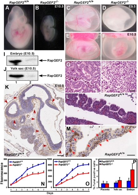
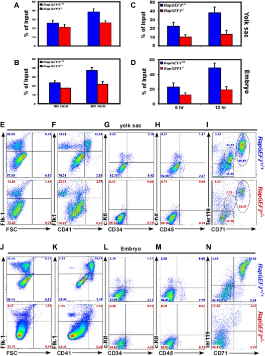
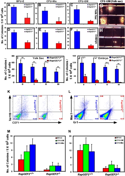
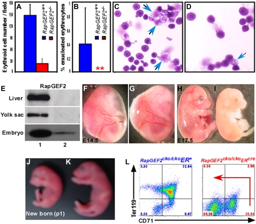
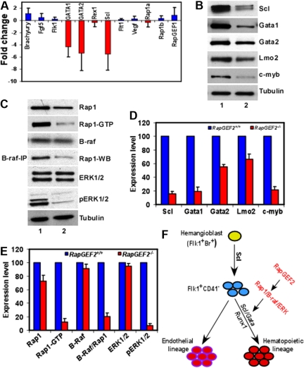
RapGEF2 is one of many guanine nucleotide exchange factors (GEFs) that specifically activate Rap1. Here, we generated RapGEF2 conditional knockout mice and studied its role in embryogenesis and fetal as well as adult hematopoietic stem cell (HSC) regulation. RapGEF2 deficiency led to embryonic lethality at ~ E11.5 due to severe yolk sac vascular defects. However, a similar number of Flk1(+) cells were present in RapGEF2(+/+) and RapGEF2(-/-) yolk sacs indicating that the bipotential early progenitors were in fact generated in the absence of RapGEF2. Further analysis of yolk sacs and embryos revealed a significant reduction of CD41 expressing cells in RapGEF2(-/-) genotype, suggesting a defect in the maintenance of definitive hematopoiesis. RapGEF2(-/-) cells displayed defects in proliferation and migration, and the in vitro colony formation ability of hematopoietic progenitors was also impaired. At the molecular level, Rap1 activation was impaired in RapGEF2(-/-) cells that in turn lead to defective B-raf/ERK signaling. Scl/Gata transcription factor expression was significantly reduced, indicating that the defects observed in RapGEF2(-/-) cells could be mediated through Scl/Gata deregulation. Inducible deletion of RapGEF2 during late embryogenesis in RapGEF2(cko/cko)ER(cre) mice leads to defective fetal liver erythropoiesis. Conversely, inducible deletion in the adult bone marrow, or specific deletion in B cells, T cells, HSCs, and endothelial cells has no impact on hematopoiesis.


Figures

Figure 1
Figure 1
Targeted disruption of the RapGEF2 gene. Strategy for generating the RapGEF2 conditional targeting vector. (A) Schematic representation of the mouse RapGEF2 genomic sequence, targeting vector, neo, cko, and ko alleles. The targeted exon 18 and its adjacent exon 19 are in red. After electroporation of the RapGEF2 targeting vector, embryonic stem (ES) cells and then the neo mice were screened by Southern blotting (B). After crossing the neo mice sequentially with the flp and β-actincre mice, the RapGEF2+/− mice were screened by Southern blotting (C). PCR strategies were used to identify the RapGEF2+/+, RapGEF2cko/+, and RapGEF2cko/cko mice (D), RapGEF2+/+, RapGEF2+/−, and RapGEF2−/− embryos and mice (E), and Cre transgenic mice (F). The RapGEF2+/− mice were fertile and apparently normal, compared with the wild-type (RapGEF2+/+) mice.
Figure 2
Figure 2
Defective yolk sac vascularization in RapGEF2−/− mice. (A-B) Morphologies of the E10.5 RapGEF2+/+ (A) and RapGEF2−/− (B) embryos. (C-D) Newly formed vitelline vessels of the E9.5 yolk sacs in RapGEF2+/+ (C) and RapGEF2−/− (D) embryos. (E-F) Vitelline vessels of the E10.5 yolk sacs in RapGEF2+/+ (E) and in RapGEF2−/− (F) embryos. The embryos were analyzed under Stemi SV11, Zeiss binocular microscope and photographed by Sony DSC-S85 digital camera. (G-H) H&E-stained transverse section of an E10.5 RapGEF2+/+ yolk sac showing blood vessels filled with red blood cells (G), and through a RapGEF2−/− yolk sac, which lacked blood vessels (H). Scale bar, 100 μm. (I-J) Northern blots showing the expression level of RapGEF2 in E10.5 RapGEF2+/+ (lane 1) and RapGEF2−/− (lane 2), embryos (I) and yolk sacs (J). (K) In situ hybridization with a RapGEF2-specific probe showing the expression pattern of RapGEF2 RNA in an E10.5 RapGEF2+/+ embryo sagittal section. Weak expression of RapGEF2 was detected throughout the embryo, but much stronger expression (indicated by red arrows) was observed in the major blood vessels. Scale bar, 500 μm. (L) H&E-stained sagittal section of an E10.5 RapGEF2+/+ yolk sac showing the yolk sac blood vessels filled with erythroblasts. Scale bar, 50 μm. (M) In situ hybridization with a RapGEF2-specific probe showing the expression pattern of RapGEF2 RNA in an E10.5 RapGEF2+/+ yolk sac. RapGEF2 was predominantly detected in the blood vessels (indicated by red arrows) of the yolk sac. Scale bar, 50 μm. (N-O) Line graph displaying the proliferation rate of RapGEF2+/+ and RapGEF2−/− E10.5 yolk sac cells (N) and embryonic cells (O) measured by AlamarBlue proliferation assay. (P) Histogram showing the percentage of Tunel-positive apoptotic cells in E10.5 RapGEF2+/+ and RapGEF2−/− placenta, yolk sac, and embryos.
Figure 3
Figure 3
CD41+ population is reduced in response to RapGEF2 deletion. (A-B) Bar graph showing the percentage of adherent RapGEF2+/+ and RapGEF2−/− E10.5 yolk sac (A) and embryonic cells (B) after 30 and 60 minutes of seeding in the fibronectin (FN)-coated plate. (C-D) Histogram displaying the in vitro migration levels of RapGEF2+/+ and RapGEF2−/− E10.5 yolk sac (C) and embryonic cells (D) after 6 and 12 hours. (E-I) Representative FACS images showing the distribution of Flk1+ (E), Flk1+CD41+ (F), c-Kit+CD34+ (G), c-Kit+CD45+ (H), and CD71+Ter119+ (I) fractions in the E10.5 RapGEF2+/+ and RapGEF2−/− yolk sacs. (J-N) Representative FACS images showing the distribution of Flk1+ (J), Flk1+CD41+ (K), c-Kit+CD34+ (L), c-Kit+CD45+ (M), and CD71+Ter119+ (N) in the E10.5 RapGEF2+/+ and RapGEF2−/− embryos.
Figure 4
Figure 4
Impaired self-renewal and differentiation of RapGEF2−/− hematopoietic progenitors in vitro. The number of BFU-E, CFU-Mix, and CFU-GM colonies formed from RapGEF2+/+ and RapGEF2−/− E10.5 yolk sac cells (A-C) or embryonic cells (D-F). The average and standard deviation were derived from 3 independent colony-formation assays. (G-H) Representative photographs showing the size of the CFU-GM colonies derived from RapGEF2+/+ (G) and RapGEF2−/− (H) E10.5 yolk sac cells. (I-J) Histograms showing the number of colonies formed in the initial plating 0′ and the subsequent replating i to iii of E10.5 yolk sac (I) or embryonic (J) cells. The average, standard deviation, and statistical significance were derived from 3 independent assays. *P < .05, **P < .005, ***P < .0005. (K-L) Flow cytometry images showing the distribution of CD71+Ter119+ (K) and Gr1+/Mac1+ (L) cell populations. (M-N) Bar graphs displaying the number of BFU-E, CFU-GM, and CFU-Mix colonies formed after plating FACS-sorted c-Kit+/CD34+ double-positive HPCs from E10.5 yolk sac (M) or embryonic cells (N). Compared with RapGEF2+/+, 5 times more number of RapGEF2−/− yolk sacs and embryos were pooled to obtain sufficient number of c-Kit+/CD34+ HPCs.
Figure 5
Figure 5
Defective fetal liver hematopoiesis in RapGEF2−/− mice. (A-B) Histogram showing the average number of nucleated (A) and enucleated (B) erythroid cells in the peripheral blood of E10.5 RapGEF2+/+ and RapGEF2−/− embryos. (C) Representative photographs displaying the cytospined and stained peripheral blood of E10.5 RapGEF2+/+ (C) and RapGEF2−/− (D) embryos. Blue arrows indicate myeloid progenitors whereas remaining cells are nucleated erythroid cells. (E) Northern blot showing the expression level of RapGEF2 in the E16.5 liver, yolk sac, and embryo of RapGEF2cko/ckoER+ (lane 1) and RapGEF2cko/ckoERcre (lane 2) mice. Females were injected with tamoxifen on day 11.5 and 13.5 of pregnancy. (F-G) Photographs showing the yolk sac blood vessels of E14.5 RapGEF2cko/ckoER+ (F) and RapGEF2cko/ckoERcre (G) embryos. (H-I) Photographs of E17.5 RapGEF2cko/ckoER+ (H) and RapGEF2cko/ckoERcre (I) embryos. The embryos were analyzed under Stemi SV11, Zeiss binocular microscope and photographed by Sony DSC-S85 digital camera. The E17.5 RapGEF2cko/ckoERcre embryos were pale, soft, fragile, and devoid of blood vessels. Timed matings were set up between the RapGEF2cko/+ERcre heterozygotes, and the pregnant females were given tamoxifen by injection to induce RapGEF2 deletion. First dose of tamoxifen was injected at E11.5, when yolk sac vascularization is mostly complete. Second dose was give at E13.5. If the induced deletion of RapGEF2 at this stage leads to embryonic lethality, we could conclude that the lethality in RapGEF2cko/ckoERcre mice was not due to yolk sac vascular defects. (J-K) Day-1 RapGEF2cko/ckoERcre (J) and RapGEF2cko/ckoER+ (K) neonates. Pregnant females were given tamoxifen by injection on days 17.5 and 19.5 of pregnancy. (L) Representative FACS images showing the distribution of the CD71+Ter119+ population in the E16.5 fetal liver cells of RapGEF2cko/ckoER+ and RapGEF2cko/ckoERcre embryos.
Figure 6
Figure 6
Deregulation of SCL/Gata transcription factors in RapGEF2−/− cells. (A) Differences in the expression level of hematopoietic marker, Brachyury, Fgf5, Flk1, Gata1, Gata2, Rex1, Scl, Flt1, and Vegf, and of Rap1a, Rap1b, and RapGEF1 in RapGEF2−/− E10.5 yolk sac cells compared with RapGEF2+/+ yolk sac cells, by Q-PCR. (B) Western blots showing the expression of the transcription factors SCL, Gata1, Gata2, and Lmo2, and the proto-oncogene c-myb in RapGEF2+/+ (lane 1) and RapGEF2−/− (lane 2) E10.5 yolk sac cells. Tubulin was used as a loading control. (C) Representative Western blots showing the level of Rap1, Rap1GTP, and B-raf, and the results of a B-raf/Rap1 pull-down assay showing the amount of B-raf-bound Rap1 (fourth panel from top), and the expression level of ERK1/2 and phosphorylated pERK1/2 in RapGEF2+/+ (lane 1) and RapGEF2−/− (lane 2) E10.5 yolk sac cells. Tubulin was used as a loading control. (D-E) The expression level of proteins (intensity of protein bands) was quantified by Imagequant software and presented as percent expression in RapGEF2−/− cells compared with RapGEF2+/+ cells, where the expression level was set as 100%. Then, the statistical analysis was shown as histograms. (F) Drawing displaying the possible role of RapGEF2 in the development of hematopoietic lineage. In this model, SCL/Gata transcription factors are required for the development of hematopoietic lineage, and RapGEF2 might regulate SCL through the Rap1/B-raf/ERK pathway.

Similar articles

Cited by

References

    1. Boettner B, Van Aelst L. Control of cell adhesion dynamics by Rap1 signaling. Curr Opin Cell Biol. 2009;21(5):684–693. - PMC - PubMed
    1. Pannekoek WJ, Kooistra MR, Zwartkruis FJ, Bos JL. Cell-cell junction formation: the role of Rap1 and Rap1 guanine nucleotide exchange factors. Biochim Biophys Acta. 2009;1788(4):790–796. - PubMed
    1. O'Keefe DD, Gonzalez-Nino E, Burnett M, et al. Rap1 maintains adhesion between cells to affect Egfr signaling and planar cell polarity in Drosophila. Dev Biol. 2009;333(1):143–160. - PMC - PubMed
    1. Retta SF, Balzac F, Avolio M. Rap1: a turnabout for the crosstalk between cadherins and integrins. Eur J Cell Biol. 2006;85(3–4):283–293. - PubMed
    1. Stork PJ, Dillon TJ. Multiple roles of Rap1 in hematopoietic cells: complementary versus antagonistic functions. Blood. 2005;106(9):2952–2961. - PMC - PubMed

Publication types

MeSH terms

Substances