Comparative Study
. 2010 Oct;5(10):1844-59.
doi: 10.2215/CJN.02210310. Epub 2010 Jul 1.

Relative role of genetic complement abnormalities in sporadic and familial aHUS and their impact on clinical phenotype

Affiliations
Comparative Study

Relative role of genetic complement abnormalities in sporadic and familial aHUS and their impact on clinical phenotype

Marina Noris et al. Clin J Am Soc Nephrol. 2010 Oct.

Abstract

Background and objectives: Hemolytic uremic syndrome (HUS) is characterized by microangiopathic hemolytic anemia, thrombocytopenia, and renal impairment. Most childhood cases are caused by Shiga toxin-producing bacteria. The other form, atypical HUS (aHUS), accounts for 10% of cases and has a poor prognosis. Genetic complement abnormalities have been found in aHUS.

Design, setting, participants, and measurements: We screened 273 consecutive patients with aHUS for complement abnormalities and studied their role in predicting clinical phenotype and response to treatment. We compared mutation frequencies and localization and clinical outcome in familial (82) and sporadic (191) cases.

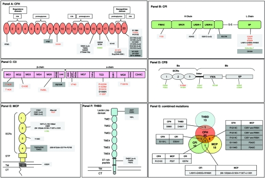
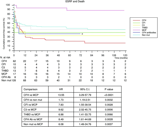
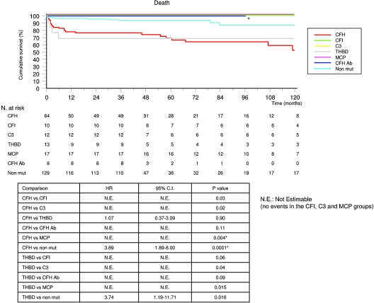
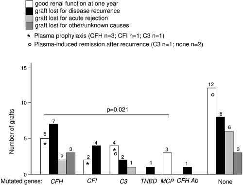
Results: In >70% of sporadic and familial cases, gene mutations, disease-associated factor H (CFH) polymorphisms, or anti-CFH autoantibodies were found. Either mutations or CFH polymorphisms were also found in the majority of patients with secondary aHUS, suggesting a genetic predisposition. Familial cases showed a higher prevalence of mutations in SCR20 of CFH and more severe disease than sporadic cases. Patients with CFH or THBD (thrombomodulin) mutations had the earliest onset and highest mortality. Membrane-cofactor protein (MCP) mutations were associated with the best prognosis. Plasma therapy induced remission in 55 to 80% of episodes in patients with CFH, C3, or THBD mutations or autoantibodies, whereas patients with CFI (factor I) mutations were poor responders. aHUS recurred frequently after kidney transplantation except for patients with MCP mutations.

Conclusions: Results underline the need of genetic screening for all susceptibility factors as part of clinical management of aHUS and for identification of patients who could safely benefit from kidney transplant.


Figures

Figure 1.
Figure 1.
Summary of CFH (A), CFI (B), C3 (C), CFB (D), MCP (E), and THBD (F) variants and of combined mutations (G) in aHUS patients from the International Registry of Recurrent and Familial HUS/TTP. Already published mutations are reported in black; new mutations are in red; and polymorphisms are in green (newly described polymorphisms are in italic). Mutations found only in familial aHUS patients are in dark gray squares, those found both in familial and sporadic cases are in gray squares, and mutations found only in sporadic cases are in light gray squares. A, B, E, and F include also mutational events in patients with combined mutations. (A) CFH: 69% of the overall independent mutations in CFH cluster in the C terminus short consensus repeat (SCR) 20. Another cluster of mutations is located in SCRs 15 to 16 (15.5%). Six mutations resulted in truncated proteins at SCR8 (n = 1), SCR15 (n = 1), SCR17 (n = 1), and SCR20 (n = 3). The aHUS-associated polymorphism in SCR16 (E936D) is marked in green. To complete CFH genetic analysis, we also screened exon 10, which produces factor H-like 1, a splice variant containing the first eight SCRs of CFH, including the complement regulatory domain. No mutations and/or polymorphisms were found. (B) CFI: 6 mutations (58%) cluster in the serine-protease domain of CFI. Of note, the intronic change 1534 + 5 G>T that was previously reported by us as a HUS-associated mutation (15) is indicated in the figure as a polymorphism because, in this report, we found this variant in a healthy control. (C) C3: the mutations are spread all over the gene; however, a hot spot is evidenced in the thioester-containing domain (TED domain) with five independent mutations (42%). (D) CFB: only one heterozygous mutation (in SCR2) has been found. Eight polymorphic variants were identified. (E) MCP: 17 independent mutations (94%) cluster in the four SCRs at the N terminus of MCP, and 55.5% are located within SCR1, confirming the importance of this region for complement regulation. The R59X and C35Y mutations were identified four and three times, respectively, suggesting that they may represent a mutational hot spot in MCP. A L139L sinonymous polymorphism has been found in MCP in a sporadic patient, but it was not found in healthy controls. This subject carries also a mutation in CFH. The amino acid syntax of MCP that takes into account the signal peptide has been adopted. (F) THBD: three independent mutational events cluster in the lectin-like domain and six cluster in the serine threonine rich (ST-rich) peptide. (G) Diagram showing the number of patients with single or combined mutations from the International Registry of Recurrent and Familial HUS/TTP is reported. Numbers of patients with mutations in CFH, MCP, CFI, and THBD alone are shown in the circles. The numbers of patients carrying combined mutations are shown in the overlapping areas; the amino acid changes are reported in the corresponding boxes.
Figure 2.
Figure 2.
Cumulative Kaplan-Meier estimates of the rates of first event (ESRF or death). The fractions of patients free of ESRF or still alive at any time point according to the presence of mutations in CFH, CFI, C3, THBD, MCP, or CFH autoantibodies or without mutations (Non mut) are shown. The MCP group was chosen as the reference group. Hazard ratios and 95% confidence intervals calculated using the Cox proportional hazards regression model are shown. P values were calculated using the log-rank test. The comparisons that were statistically significant after Bonferroni correction are shown in the table. °Follow-up < 120 months.
Figure 3.
Figure 3.
Cumulative Kaplan-Meier estimates of the rates of death. Fractions of patients still alive at any time point according to the presence of mutations in CFH, CFI, C3, THBD, MCP, or CFH autoantibodies or without mutations (Non mut) are shown. The CFH and THBD groups were chosen as reference groups. When feasible, hazard ratios and 95% confidence intervals were calculated by means of Cox proportional hazards regression model and shown in the figure. P values were calculated by means of the log-rank test after Bonferroni's correction. The most relevant comparisons are shown in the table. *Statistically significant after Bonferroni correction. °Follow-up < 120 months.
Figure 4.
Figure 4.
Outcome of kidney transplantation. The outcome at 1 year of 64 transplanted kidneys in genotyped patients of the International Registry of HUS/TTP is reported, according to the absence or presence of a mutation. Plasma prophylaxis was used in three patients with CFH mutation, in one patient with C3 mutation, and in one patient with CFI mutation (*), all with good outcomes. Plasma was given to treat aHUS recurrences in 20 grafts. Remission was achieved in only three (°). The numbers of transplanted organs are shown above each column.

Similar articles

Cited by

References

    1. Ruggenenti P, Noris M, Remuzzi G: Thrombotic microangiopathy, hemolytic uremic syndrome, and thrombotic thrombocytopenic purpura. Kidney Int 60: 831–846, 2001 - PubMed
    1. Besbas N, Karpman D, Landau D, Loirat C, Proesmans W, Remuzzi G, Rizzoni G, Taylor CM, Van de Kar N, Zimmerhackl LB: A classification of hemolytic uremic syndrome and thrombotic thrombocytopenic purpura and related disorders. Kidney Int 70: 423–431, 2006 - PubMed
    1. Scheiring J, Andreoli SP, Zimmerhackl LB: Treatment and outcome of Shiga-toxin-associated hemolytic uremic syndrome (HUS). Pediatr Nephrol 23: 1749–1760, 2008 - PMC - PubMed
    1. Noris M, Remuzzi G: Atypical hemolytic-uremic syndrome. N Engl J Med 361: 1676–1687, 2009 - PubMed
    1. Kavanagh D, Goodship TH: Update on evaluating complement in hemolytic uremic syndrome. Curr Opin Nephrol Hypertens 16: 565–571, 2007 - PubMed

Publication types

MeSH terms