. 2010 Jul 1;466(7302):62-7.
doi: 10.1038/nature09130.

Chromatin regulation by Brg1 underlies heart muscle development and disease

Affiliations

Chromatin regulation by Brg1 underlies heart muscle development and disease

Calvin T Hang et al. Nature. .

Erratum in

  • Nature. 2011 Jul 28;475(7357):532

Abstract

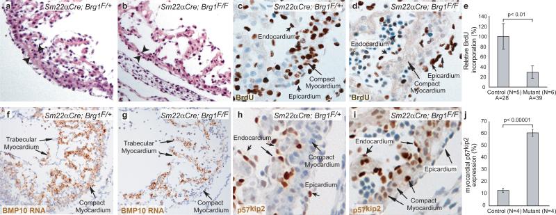
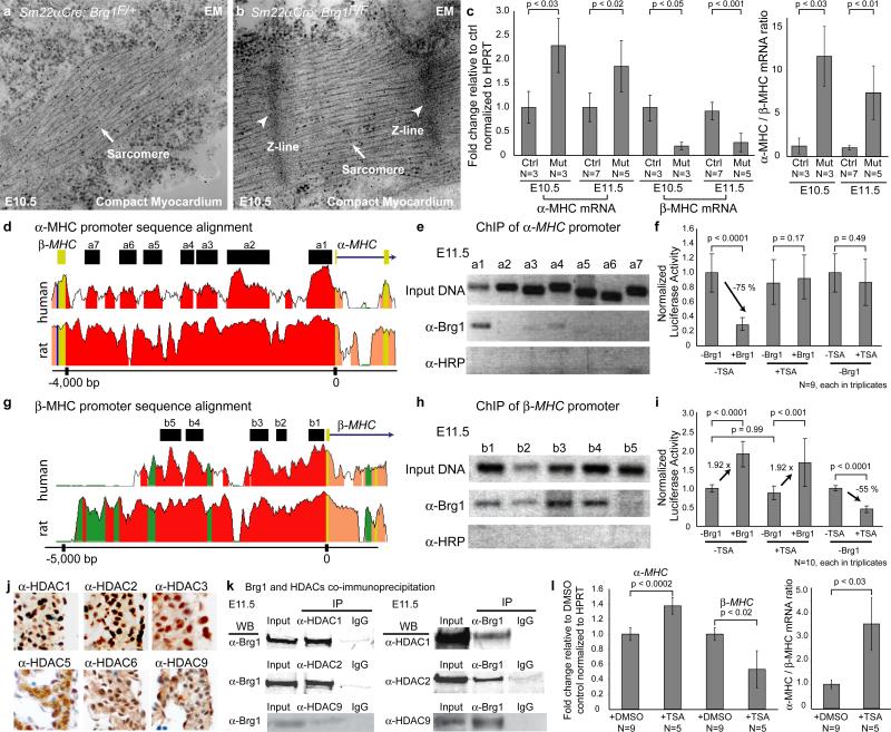
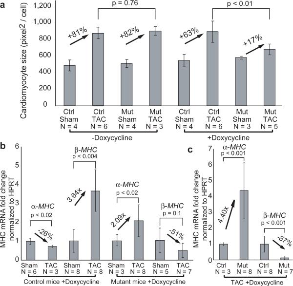
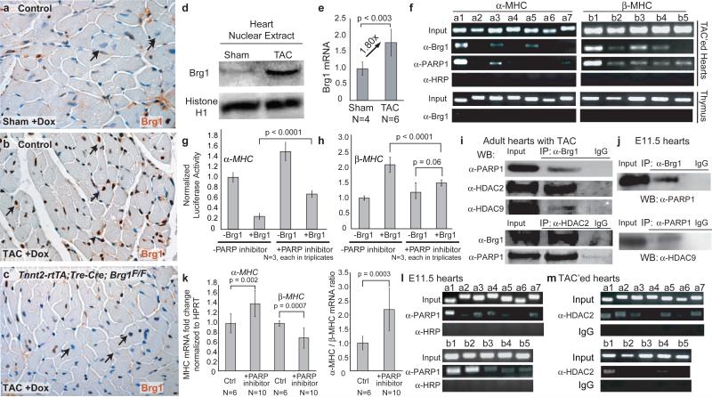
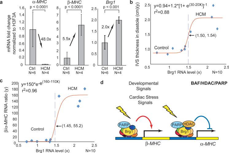
Cardiac hypertrophy and failure are characterized by transcriptional reprogramming of gene expression. Adult cardiomyocytes in mice primarily express alpha-myosin heavy chain (alpha-MHC, also known as Myh6), whereas embryonic cardiomyocytes express beta-MHC (also known as Myh7). Cardiac stress triggers adult hearts to undergo hypertrophy and a shift from alpha-MHC to fetal beta-MHC expression. Here we show that Brg1, a chromatin-remodelling protein, has a critical role in regulating cardiac growth, differentiation and gene expression. In embryos, Brg1 promotes myocyte proliferation by maintaining Bmp10 and suppressing p57(kip2) expression. It preserves fetal cardiac differentiation by interacting with histone deacetylase (HDAC) and poly (ADP ribose) polymerase (PARP) to repress alpha-MHC and activate beta-MHC. In adults, Brg1 (also known as Smarca4) is turned off in cardiomyocytes. It is reactivated by cardiac stresses and forms a complex with its embryonic partners, HDAC and PARP, to induce a pathological alpha-MHC to beta-MHC shift. Preventing Brg1 re-expression decreases hypertrophy and reverses this MHC switch. BRG1 is activated in certain patients with hypertrophic cardiomyopathy, its level correlating with disease severity and MHC changes. Our studies show that Brg1 maintains cardiomyocytes in an embryonic state, and demonstrate an epigenetic mechanism by which three classes of chromatin-modifying factors-Brg1, HDAC and PARP-cooperate to control developmental and pathological gene expression.


Figures

Figure 1
Figure 1. Brg1 promotes myocardial proliferation
a, b, H&E sections of E10.5 compact myocardium, whose thickness is denoted by arrowheads. c, d, BrdU immunostaining of E10.5 compact myocardium. e, BrdU incorporation quantitation. A: number of areas examined. p-value: Student's t-test. Error bar: standard deviation. f, g, BMP10 in situ hybridization of E10.5 hearts. h, i, p57kip2 immunostaining of E10.5 hearts. j, p57kip2 quantitation. p-value: Student's t-test. Error bar: standard deviation.
Figure 2
Figure 2. Brg1 suppresses myocardial differentiation
a, b, EM of the compact myocardium of E10.5 embryos. c, Quantitative RT-PCR of ventricular α- and β-MHC at E10.5 and E11.5. Ctrl: control. Mut: Sm22αCre;Brg1F/F. p-value: Student's t-test. Error bar: standard deviation. d, Sequence alignment of the α-MHC locus from mouse, human, and rat. Peak heights indicate degree of sequence homology. Black boxes (a1-a7) are regions of high sequence homology and further analyzed by ChIP. Red: promoter elements. Salmon: introns. Yellow: untranslated regions. e, PCR of Brg1-immunoprecipitated chromatin from E11.5 hearts. α-HRP: anti-horse radish peroxidase antibody. f, Luciferase reporter assay of the proximal α-MHC promoter (-462 to +192) in SW13 cells. p-value: Student's t-test. Error bar: standard deviation. g, Sequence alignment of the β-MHC locus from mouse, human, and rat. Black boxes (b1-b5) are regions of high sequence homology and further analyzed by ChIP. Green: transposons/simple repeats. h, PCR analysis of Brg1-immunoprecipitated chromatin from E11.5 hearts. i, Luciferase reporter assays of the β-MHC proximal promoter (-835 to +222) in SW13 cells. p-value: Student's t-test. Error bar: standard deviation. j, Immunostaining of HDAC1, 2, 3, 5, 6 and 9 (brown) in E11.5 hearts. k, Co-immunoprecipitation of Brg1 with HDAC1, 2 and 9 in E11.5 hearts. l, Quantitative RT-PCR of α- and β-MHC of cultured embryos treated with DMSO or TSA. p-value: Student's t-test. Error bar: standard deviation.
Figure 3
Figure 3. Brg1 is required for cardiac hypertrophy
a, Cardiomyocyte size quantitation. Ctrl: control. Mut: Tnnt2-rtTA;Tre-Cre;Brg1F/F. p-value: Student's t-test. Error bar: standard deviation. b, c, Quantitative RT-PCR of α- and β-MHC in cardiac ventricles of doxycycline-treated control and Tnnt2-rtTA;Tre-Cre;Brg1F/F mice 4 weeks after sham/TAC operation. p-value: Student's t-test. Error bar: standard deviation.
Figure 4
Figure 4. MHC regulation by Brg1, PARP and HDAC
a, b, c, Brg1 immunostaining in ventricular myocardium of doxycycline-treated control and Tnnt2-rtTA;Tre-Cre;Brg1F/F mice 1 week after sham/TAC operation. Arrows: cardiomyocyte nuclei. d, Brg1 immunoblot of cardiac nuclear extracts from wildtype mice 2 weeks after TAC. e, Quantitative RT-PCR of Brg1 mRNA in wildtype mice 2 weeks after TAC. p-value: Student's t-test. Error bar: standard deviation. f, PCR of Brg1- and PARP1- immunoprecipitated chromatin from thymus and adult hearts 2 weeks after TAC. g, h, Luciferase reporter assays of α-MHC (g) and β-MHC (h) promoter in SW13 cells with PARP inhibition. p-value: Student's t-test. Error bar: standard deviation. i, j, Co-immunoprecipitation of Brg1, PARP1, HDAC2 and 9 in TAC-treated adult hearts (i) and in E11.5 hearts (j). k, Quantitative RT-PCR of α- and β-MHC of PJ34-treated cultured embryos. p-value: Student's t-test. Error bar: standard deviation. l, PCR of PARP1- immunoprecipitated chromatin from E11.5 hearts. m, PCR of HDAC2- immunoprecipitated chromatin from adult hearts 2 weeks after TAC.
Figure 5
Figure 5. Brg1 activation in human cardiomyopathy
a, Quantitative RT-PCR of α-MHC, β-MHC, and Brg1 expression in normal and HCM subjects. p-value: Student's t-test. Error bar: standard deviation. b, IVSd (y) plotted against the Brg1 RNA level (x). Red: regression curve. e: the base of natural logarithm (~2.718). Arrow and dashed line: the inflection point. c, The β /α-MHC RNA ratio (y) plotted against the Brg1 RNA level (x). d, Model of developmentally-activated and stress-induced assembly of BAF/HDAC/PARP complexes on the α-MHC, and BAF/PARP complex on the β-MHC promoter.

Similar articles

Cited by

References

    1. van Rooij E, et al. Control of stress-dependent cardiac growth and gene expression by a microRNA. Science. 2007;316:575–9. - PubMed
    1. Herron TJ, McDonald KS. Small amounts of alpha-myosin heavy chain isoform expression significantly increase power output of rat cardiac myocyte fragments. Circ Res. 2002;90:1150–2. - PubMed
    1. Krenz M, Robbins J. Impact of beta-myosin heavy chain expression on cardiac function during stress. J Am Coll Cardiol. 2004;44:2390–7. - PubMed
    1. James J, et al. Forced expression of alpha-myosin heavy chain in the rabbit ventricle results in cardioprotection under cardiomyopathic conditions. Circulation. 2005;111:2339–46. - PMC - PubMed
    1. Miyata S, Minobe W, Bristow MR, Leinwand LA. Myosin heavy chain isoform expression in the failing and nonfailing human heart. Circ Res. 2000;86:386–90. - PubMed

Publication types

MeSH terms