. 2010 Jul 1;466(7302):113-7.
doi: 10.1038/nature09114.

Genome-wide association study in alopecia areata implicates both innate and adaptive immunity

Affiliations

Genome-wide association study in alopecia areata implicates both innate and adaptive immunity

Lynn Petukhova et al. Nature. .

Abstract

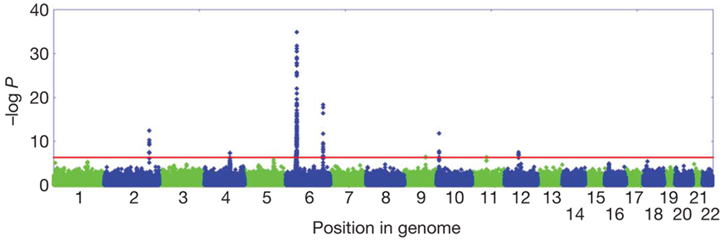
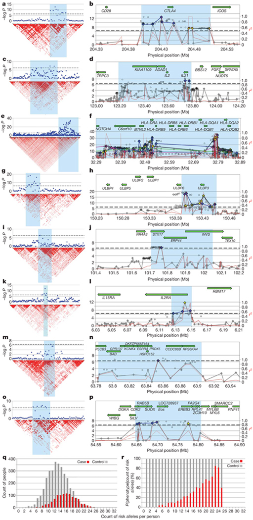
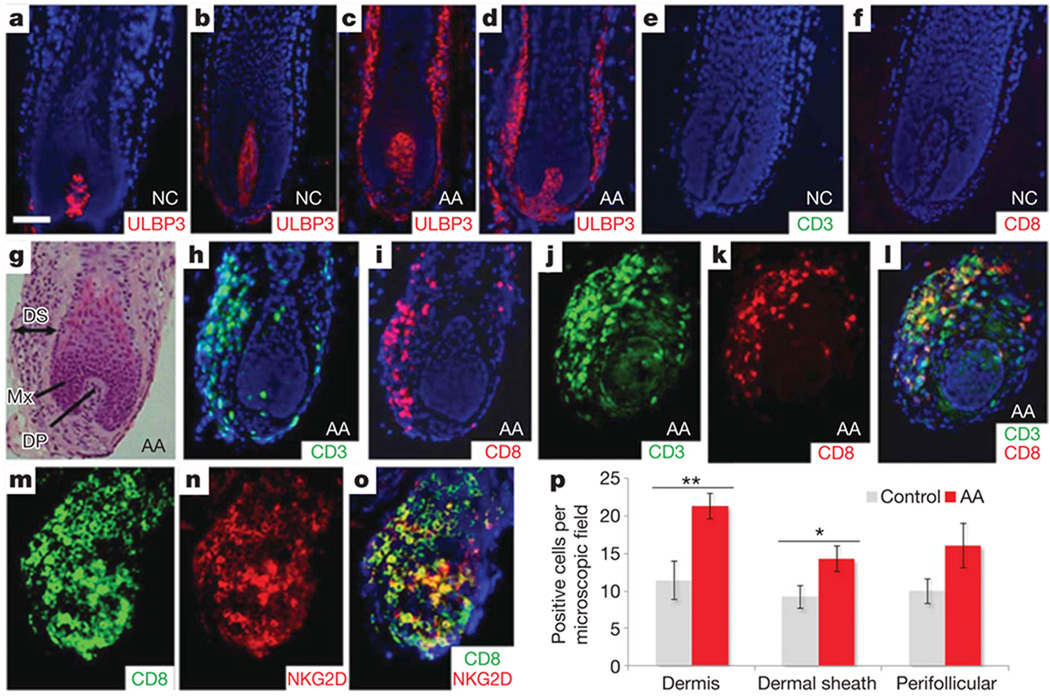
Alopecia areata (AA) is among the most highly prevalent human autoimmune diseases, leading to disfiguring hair loss due to the collapse of immune privilege of the hair follicle and subsequent autoimmune attack. The genetic basis of AA is largely unknown. We undertook a genome-wide association study (GWAS) in a sample of 1,054 cases and 3,278 controls and identified 139 single nucleotide polymorphisms that are significantly associated with AA (P <or= 5 x 10(-7)). Here we show an association with genomic regions containing several genes controlling the activation and proliferation of regulatory T cells (T(reg) cells), cytotoxic T lymphocyte-associated antigen 4 (CTLA4), interleukin (IL)-2/IL-21, IL-2 receptor A (IL-2RA; CD25) and Eos (also known as Ikaros family zinc finger 4; IKZF4), as well as the human leukocyte antigen (HLA) region. We also find association evidence for regions containing genes expressed in the hair follicle itself (PRDX5 and STX17). A region of strong association resides within the ULBP (cytomegalovirus UL16-binding protein) gene cluster on chromosome 6q25.1, encoding activating ligands of the natural killer cell receptor NKG2D that have not previously been implicated in an autoimmune disease. By probing the role of ULBP3 in disease pathogenesis, we also show that its expression in lesional scalp from patients with AA is markedly upregulated in the hair follicle dermal sheath during active disease. This study provides evidence for the involvement of both innate and acquired immunity in the pathogenesis of AA. We have defined the genetic underpinnings of AA, placing it within the context of shared pathways among autoimmune diseases, and implicating a novel disease mechanism, the upregulation of ULBP ligands, in triggering autoimmunity.


Figures

Figure 1
Figure 1. Manhattan plot of the joint analysis of the discovery GWAS and the replication GWAS
Results are plotted as negative log-transformed P values from a genotypic association test controlled for residual population stratification as a function of the position in the genome. Odd chromosomes are in green and even chromosomes in blue. Eight genomic regions contain SNPs that exceed the genome-wide significance threshold of 5 × 10⁻⁷ (red line).
Figure 2
Figure 2. LD structure and haplotype organization of the implicated regions from GWAS
ap, In all graphs the genome-wide significance threshold (5 × 10⁻⁷) is indicated by a black dotted line. Results from the eight regions are aligned with LD maps (a, c, e, g, i, k,m, o) and transcript maps (b, d, f, h, j, l, n, p): chromosomes 2q33 (a, b), 4q26–27 (c, d), 6p21.3 (e, f), 6q25.1 (g, h), 9q31.1 (i, j), 10p15–p16 (k, l), 11q13 (m, n) and 12q13 (o, p). For the plots with the LD maps, red indicates high LD as measured by D′. For the plots with the transcript maps, results for imputed SNPs are indicated by open circles. Typed SNPs that do not reach significance are in grey; significantly associated typed SNPs are in colour, coded by the risk haplotypes. For example in b, conditioning on any of the blue SNPs will decrease evidence for association of the other blue SNPs but will not affect evidence of any of the yellow-coded SNPs. On chromosome 6p in the HLA, significantly associated SNPs can be organized into at least five distinct haplotypes (blue, green, yellow, pink and light blue). The red lines show pairwise LD as measured by r2, for the most significant SNP in each haplotype, and define the LD block that is demonstrating association. q, r, The cumulative effect of risk haplotypes is indicated by the distribution of the genetic liability index in cases and controls. We chose the most significantly associated SNP from each haplotype to serve as a proxy for the haplotype, and show in q that the distribution of independent genetic risk factors changes as a function of phenotype, with an average of 13 risk alleles found in controls (grey) and 16 found in cases (red). As the number of risk alleles in an individual increases, the proportion affected by AA increases. The conditional probability of phenotype given count of risk alleles is shown in r (AA in red, control in grey).
Figure 3
Figure 3. ULBP3 expression and immune cell infiltration of AA hair follicles
a, b, Low levels of expression of ULBP3 in the dermal papilla of hair follicles from two unrelated, unaffected individuals. c, d, Massive upregulation of ULBP3 expression in the dermal sheath of hair follicles from two unrelated patients with AA in the early stages of disease. e, f, Absence of immune infiltration in two control hair follicles. g, Haematoxylin and eosin staining of AA hair follicle. DS, dermal sheath; Mx, matrix; DP, dermal papilla. h, i, Immunofluorescence analysis using CD3 (h) and CD8 (i) cell-surface markers for T-cell lineages. There is a marked inflammatory infiltrate in the dermal sheath of two affected AA hair follicles. jl, Double-immunofluorescence analysis with anti-CD3 (j) and anti-CD8 (k) antibodies. l, The merged image clearly shows infiltration of CD3+CD8+ T cells in the dermal sheath of the AA hair follicle. Panels d and gl are serial sections of the same hair follicle of an affected individual. Counterstaining with 4′,6-diamidino-2-phenylindole is shown in blue (af, h, i, l). Scale bar, 50 µm. AA, alopecia areata patients; NC, normal control individuals. mo, Double immunostainings with anti-CD8 (m) and anti-NKG2D (n) antibodies revealed that most NKG2D+ cells co-expressed CD8+; o, merged image. p, Quantification of immunohistochemical staining (positive cells per microscope field at a magnification of × 200) for ULBP3 in 16 patients with AA and in 7 controls showed a significantly increased number of ULBP3+ cells in the dermis and dermal sheath in patients with AA compared with control skin. In addition, positive cells were also upregulated in perifollicular regions in AA samples, although this was not statistically significant. Data were analysed by Mann–Whitney test for unpaired samples and are expressed as means ± s.e.m.; asterisk, P < 0.05; two asterisks, P < 0.01.

Similar articles

Cited by

References

    1. Cooper GS, Bynum ML, Somers EC. Recent insights in the epidemiology of autoimmune diseases: improved prevalence estimates and understanding of clustering of diseases. J. Autoimmun. 2009;33:197–207. - PMC - PubMed
    1. Safavi KH, Muller SA, Suman VJ, Moshell AN, Melton LJ., III Incidence of alopecia areata in Olmsted County, Minnesota, 1975 through 1989. Mayo Clin. Proc. 1995;70:628–633. - PubMed
    1. Jelinek JE. Sudden whitening of the hair. Bull. N. Y. Acad. Med. 1972;48:1003–1013. - PMC - PubMed
    1. Gilhar A, Paus R, Kalish RS. Lymphocytes, neuropeptides, and genes involved in alopecia areata. J. Clin. Invest. 2007;117:2019–2027. - PMC - PubMed
    1. Gilhar A, et al. Transfer of alopecia areata in the human scalp graft/Prkdc(scid) (SCID) mouse system is characterized by a TH1 response. Clin. Immunol. 2003;106:181–187. - PubMed

Publication types

MeSH terms