. 2010 Jul 1;466(7302):133-7.
doi: 10.1038/nature09161.

Human melanoma-initiating cells express neural crest nerve growth factor receptor CD271

Affiliations

Human melanoma-initiating cells express neural crest nerve growth factor receptor CD271

Alexander D Boiko et al. Nature. .

Erratum in

  • Nature. 2011 Feb 17;470(7334):424

Abstract

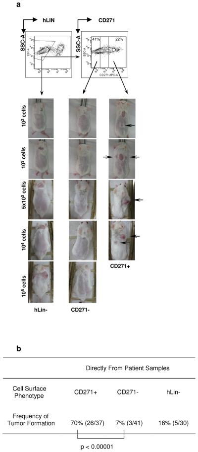
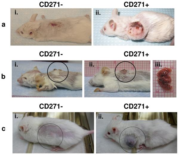
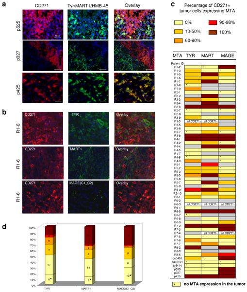
The question of whether tumorigenic cancer stem cells exist in human melanomas has arisen in the last few years. Here we show that in melanomas, tumour stem cells (MTSCs, for melanoma tumour stem cells) can be isolated prospectively as a highly enriched CD271(+) MTSC population using a process that maximizes viable cell transplantation. The tumours sampled in this study were taken from a broad spectrum of sites and stages. High-viability cells isolated by fluorescence-activated cell sorting and re-suspended in a matrigel vehicle were implanted into T-, B- and natural-killer-deficient Rag2(-/-)gammac(-/-) mice. The CD271(+) subset of cells was the tumour-initiating population in 90% (nine out of ten) of melanomas tested. Transplantation of isolated CD271(+) melanoma cells into engrafted human skin or bone in Rag2(-/-)gammac(-/-) mice resulted in melanoma; however, melanoma did not develop after transplantation of isolated CD271(-) cells. We also show that in mice, tumours derived from transplanted human CD271(+) melanoma cells were capable of metastatsis in vivo. CD271(+) melanoma cells lacked expression of TYR, MART1 and MAGE in 86%, 69% and 68% of melanoma patients, respectively, which helps to explain why T-cell therapies directed at these antigens usually result in only temporary tumour shrinkage.


Figures

Figure 1
Figure 1. Isolation of melanoma tumor stem cells (MTSC) expressing CD271P75(NGFR) from melanoma patients
a, Representative contour plot FACS gating sequence leading to purification of live, hLin- (CD45−/CD2−/CD3−/ CD31−), CD271+ and C271− cells from Mel114 patient sample; CD271+ but not CD271− melanoma cells induce tumors upon intradermal injection in 30% matrigel into B-, T- and NK cell deficient Rag2−/− γc−/− (RG) mice after 28-32 weeks. b, Summary Table of tumor formation frequencies by CD271+ and CD271− human melanoma cells isolated from all patients.
Figure 2
Figure 2. MTSCs induce tumors in humanized mouse models
a, Humanized RG mice that contained skin grafts from the same healthy donor were used to assess the tumorigenic potential of CD271+ cells. Dermal melanoma xenograft (P0) from a primary patient (Mel43) was used to purify live, mLin-, CD271+ and CD271− melanoma cells by FACS and injected into separate human skin grafts on RG mice from the same healthy donor. (i) human skin graft of RG mice 28 weeks after injection of 2×10⁴ CD271− cells and (ii) melanoma formation in human skin graft of RG mice 28 weeks after injection of 2×10⁴ of CD271+ cells; b, Surgically resected primary melanoma from mel826 patient was used to isolate live, hLin-, CD271+ and CD271− cells by FACS and injected into separate human skin grafts on RG mice from the same healthy donor; (i) human skin graft 16 weeks after injection of 2×10⁴ CD271− cells; (ii)-(iii) melanoma tumor formation and lung metastasis 16 weeks after injection of 6×10³ CD271+ cells; c, Surgically resected melanoma adjacent to the patella from the patient mel210 was used to purify live, hLin-, CD271+ and CD271− melanoma cells by FACS; 103 cells of each highly purified fraction were injected separately into two NSG mice subcutaneously near the grafted human bone fragment (i) human bone graft of NSG mice 20 weeks after injection of 103 CD271− cells and (ii) 103 CD271+ cells.
Figure 3
Figure 3. Most melanomas contain CD271+ tumor cells that either completely or partially lack expression of MTAs
a. Immunofluorescent analysis of CD271, Tyr/MART1/HMB-45 expression in tissue sections of melanoma patients Mel525, Mel327 and Mel 425; b, Representative images of immunofluorescent analysis of CD271, TYR, MART1 and MAGE (C1-C2) expression in tissue cores of melanoma patient from tissue array. c, Table indicating expression of melanoma tumor antigens (MTAs) TYR, MART1 and MAGE C1-C2 in CD271+ cells of each patient's tumor. Colour bars indicate percentage range of CD271+ cells that expressed MTA; grey bars indicate that expression of CD271 and MTA was not detected during analysis of the tumor core. d, Stacked bar graph indicating proportion of melanoma patients with ranges of MTA positivity of CD271+ cells.

Comment in

References

    1. Quintana E, et al. Efficient tumour formation by single human melanoma cells. Nature. 2008;456:593–8. - PMC - PubMed
    1. Weissman I. Stem cell research: paths to cancer therapies and regenerative medicine. Jama. 2005;294:1359–66. - PubMed
    1. Al-Hajj M, Wicha MS, Benito-Hernandez A, Morrison SJ, Clarke MF. Prospective identification of tumorigenic breast cancer cells. Proc Natl Acad Sci U S A. 2003;100:3983–8. - PMC - PubMed
    1. Jamieson CH, et al. Granulocyte-macrophage progenitors as candidate leukemic stem cells in blast-crisis CML. N Engl J Med. 2004;351:657–67. - PubMed
    1. Singh SK, et al. Identification of a cancer stem cell in human brain tumors. Cancer Res. 2003;63:5821–8. - PubMed

Publication types

MeSH terms