. 2010 Jul 1;466(7302):138-42.
doi: 10.1038/nature09140.

SCF(Cyclin F) controls centrosome homeostasis and mitotic fidelity through CP110 degradation

Affiliations

SCF(Cyclin F) controls centrosome homeostasis and mitotic fidelity through CP110 degradation

Vincenzo D'Angiolella et al. Nature. .

Abstract

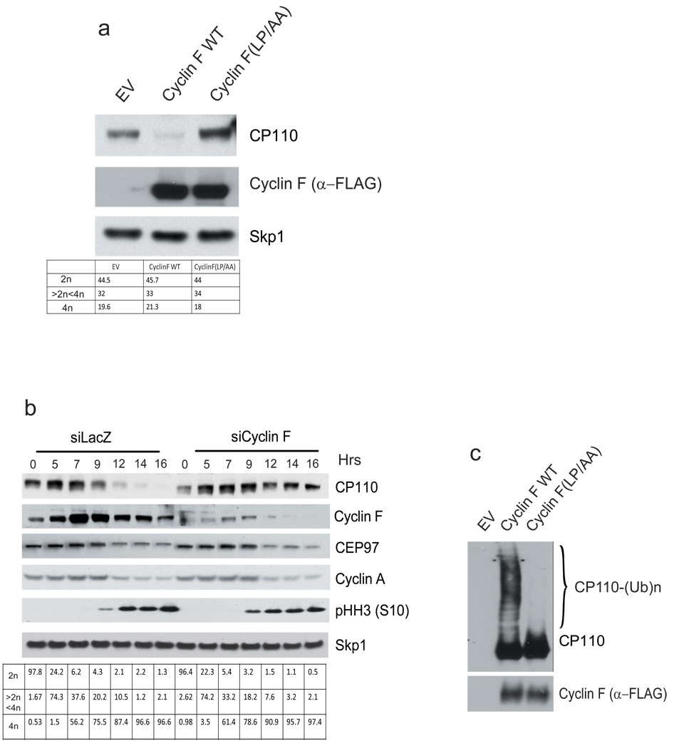
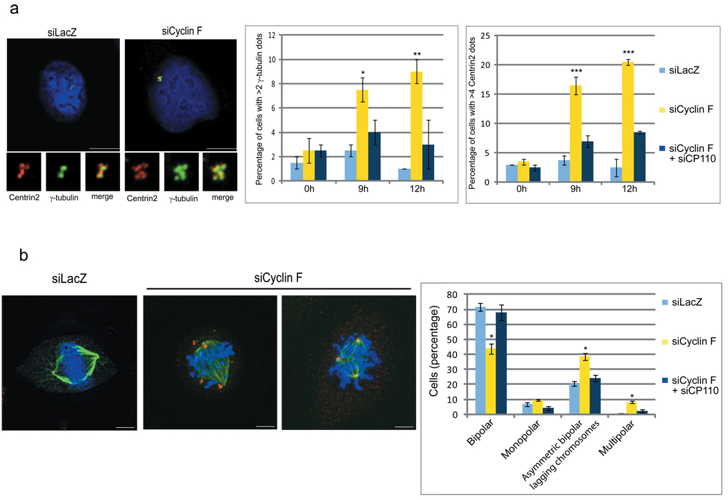
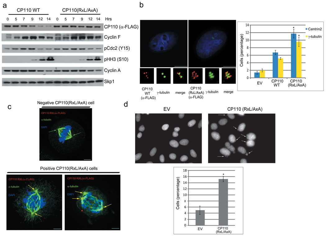
Generally, F-box proteins are the substrate recognition subunits of SCF (Skp1-Cul1-F-box protein) ubiquitin ligase complexes, which mediate the timely proteolysis of important eukaryotic regulatory proteins. Mammalian genomes encode roughly 70 F-box proteins, but only a handful have established functions. The F-box protein family obtained its name from Cyclin F (also called Fbxo1), in which the F-box motif (the approximately 40-amino-acid domain required for binding to Skp1) was first described. Cyclin F, which is encoded by an essential gene, also contains a cyclin box domain, but in contrast to most cyclins, it does not bind or activate any cyclin-dependent kinases (CDKs). However, like other cyclins, Cyclin F oscillates during the cell cycle, with protein levels peaking in G2. Despite its essential nature and status as the founding member of the F-box protein family, Cyclin F remains an orphan protein, whose functions are unknown. Starting from an unbiased screen, we identified CP110, a protein that is essential for centrosome duplication, as an interactor and substrate of Cyclin F. Using a mode of substrate binding distinct from other F-box protein-substrate pairs, CP110 and Cyclin F physically associate on the centrioles during the G2 phase of the cell cycle, and CP110 is ubiquitylated by the SCF(Cyclin F) ubiquitin ligase complex, leading to its degradation. siRNA-mediated depletion of Cyclin F in G2 induces centrosomal and mitotic abnormalities, such as multipolar spindles and asymmetric, bipolar spindles with lagging chromosomes. These phenotypes were reverted by co-silencing CP110 and were recapitulated by expressing a stable mutant of CP110 that cannot bind Cyclin F. Finally, expression of a stable CP110 mutant in cultured cells also promotes the formation of micronuclei, a hallmark of chromosome instability. We propose that SCF(Cyclin F)-mediated degradation of CP110 is required for the fidelity of mitosis and genome integrity.


Figures

Figure 1
Figure 1. Cyclin F and CP110 interact and colocalize to the centrosomes
a, HeLa cells were synchronized at G1/S using a double-thymidine block before release into fresh medium. Cells were collected at the indicated times, lysed, immunoprecipitated with anti-Cyclin F antibody, and immunoblotted as indicated. Last lane shows immunoprecipitation pre-incubated with the antigenic peptide. Left panel show 10% of the material used for immunoprecipitations (input). b, U-2OS cells were synchronized as in (a), fixed, and incubated with an anti-Cyclin F antibody (red) and anti-γ-tubulin antibody (green). The panel shows staining after pre-incubation with the antigenic peptide. DNA was stained with DAPI. Insets show magnified views of the centrosomes indicated by arrows. Scale bar = 10 µM. The table shows the percentage of cells with centrosomal Cyclin F (where 100% was the total cells staining positive for nuclear Cyclin F) at different times after the release from the double-thymidine block (n = ~50 per time point). c, Cyclin F and CP110 colocalize to the centrioles. U-2OS cells were transfected with GFP-CP110 and Cherry-Cyclin F. In merged images, yellow shows colocalization of CP110 and Cyclin F.
Figure 2
Figure 2. CP110 is targeted for ubiquitylation and degradation by SCFCyclin F during the G2 phase of the cell cycle
a, U-2OS cells were transfected with an empty vector (EV), FLAG-tagged Cyclin F, or FLAG-tagged Cyclin F(LP/AA). Twenty-four hours after transfection, cells were collected, lysed, and immunoblotted as indicated. DNA content was monitored by flow cytometry. b, HeLa cells were transfected with short interfering RNAs (siRNAs) to either LacZ or Cyclin F (oligo #2). Cells synchronized as in (1a) were then collected at the indicated times, lysed, and immunoblotted as indicated. DNA content was monitored by flow cytometry. c, HEK-293T cells were transfected as in (2a), lysed, immunoprecipitated with anti-FLAG resin, and used in a ubiquitylation assay.
Figure 3
Figure 3. Cyclin F silencing induces centrosome and mitotic aberrations
a, U-2OS cells were transfected with siRNAs to a LacZ, Cyclin F, or both Cyclin F and CP110, synchronized as in (1a), fixed at the indicated times after release from the block, and incubated with anti-Centrin 2 antibody (red) and anti-γ-tubulin antibody (green). DNA was stained with DAPI. Insets show magnified views of centrosomes. Scale bar = 10 µM. The graphs on the right show quantification of three experiments. Error bars indicate +/−SD. *= p=0.003; **= p=0.001; ***= p<0.001. b, Experiments were performed as in (a), except that cells were collected 14 hours after release from the block and stained with anti-Centrin 2 antibody (red) and anti-α-tubulin antibody (green). Scale bar = 5 µM. The graphs on the right show the percentages of cells with various abnormal mitotic phenotypes. Error bars indicate +/−SD. *= p<0.001.
Figure 4
Figure 4. The failure to degrade CP110 causes centrosome and mitotic defects
a, HeLa cells were transfected with FLAG-CP110 or FLAG-CP110(RxL/AxA), synchronized at G1/S as in (1a), collected at the indicated times, lysed, and immunoblotted as indicated. b, U-2OS cells were transfected with FLAG-CP110 or FLAG-CP110(RxL/AxA), collected 48 hours after transfection, and stained with anti-FLAG antibody to visualize CP110 (red) and anti-γ-tubulin (green) antibody. Insets show magnified views of centrosomes. Scale bar = 10 µM. The graphs on the right show quantification of cells with excess Centrin 2 and γ-tubulin dots. Error bars indicate +/−SD. *= p<0.001 (n=3). c, Experiments were performed as in (b), except that cells were transfected with only FLAG-CP110(RxL/AxA), collected 14 hours after release from the block, and stained with anti-α-tubulin antibody (green) and anti-FLAG antibody to visualize CP110-positive cells (red). The yellow in merged images shows colocalization of CP110 and α-tubulin (yellow arrows). Green arrows show spindles negative for CP110. CP110 negative and CP110 positive cells from the same coverslip are shown. Scale bar = 5 µM. d, U-2OS cells were transfected with an empty vector (EV) or FLAG-CP110(RxL/AxA), fixed, and stained with DAPI to visualize the DNA. Micronuclei are highlighted by arrows. The graph on the bottom shows quantification of cells containing micronuclei. Error bars indicate +/−SD. *= p<0.0001.

Similar articles

Cited by

References

    1. Cardozo T, Pagano M. The SCF Ubiquitin Ligase: Insights into a Molecular Machine. Nat Rev Mol Cell Biol. 2004;5:739–751. - PubMed
    1. Petroski MD, Deshaies RJ. Function and regulation of cullin-RING ubiquitin ligases. Nat Rev Mol Cell Biol. 2005;6:9–20. - PubMed
    1. Jin J, et al. Systematic analysis and nomenclature of mammalian F-box proteins. Genes Dev. 2004;18:2573–2580. - PMC - PubMed
    1. Skaar JR, D'Angiolella V, Pagan JK, Pagano M. SnapShot: F Box Proteins II. Cell. 2009;137:1358–1358. e1351. - PubMed
    1. Bai C, et al. Skp1 connects cell cycle regulators to the ubiquitin proteolysis machinery through a novel motif, the F-box. Cell. 1996;86:263–274. - PubMed

Publication types

MeSH terms