. 2011 Jan;18(1):80-9.
doi: 10.1038/cdd.2010.81. Epub 2010 Jul 2.

Blebbing confers resistance against cell lysis

Affiliations

Blebbing confers resistance against cell lysis

E B Babiychuk et al. Cell Death Differ. 2011 Jan.

Abstract

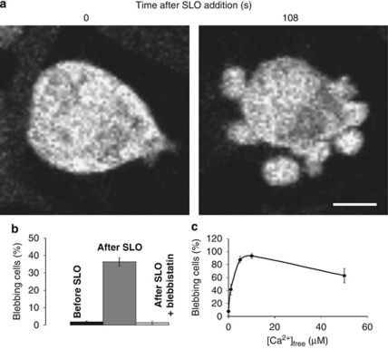
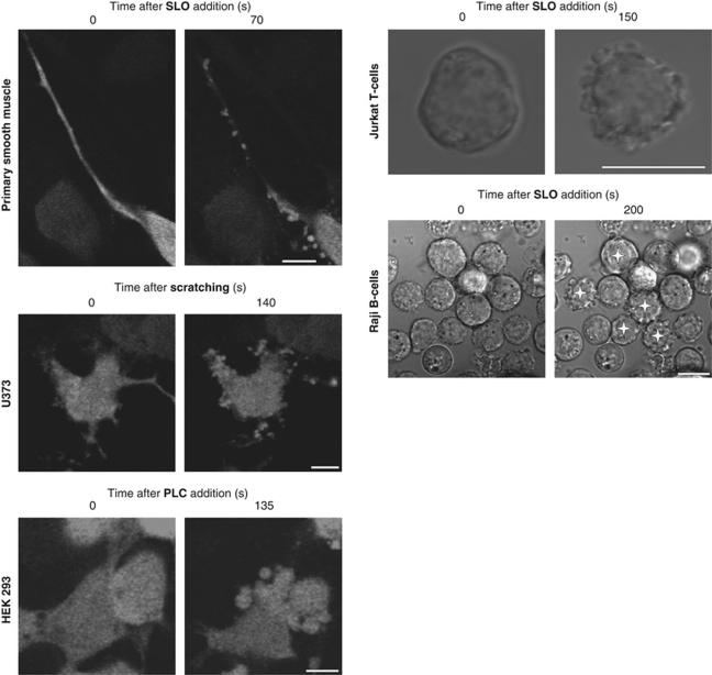
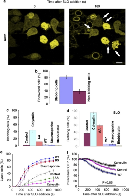
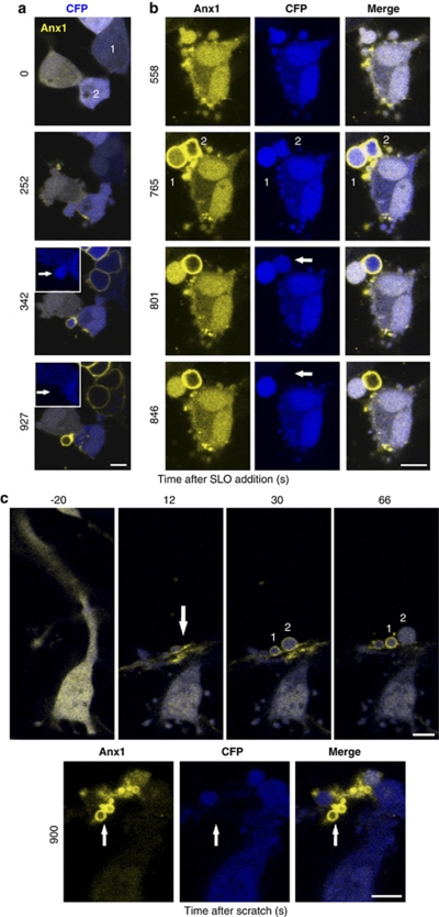
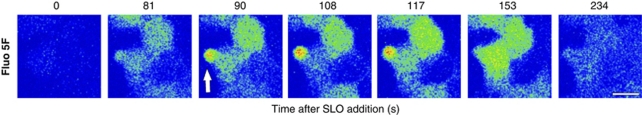
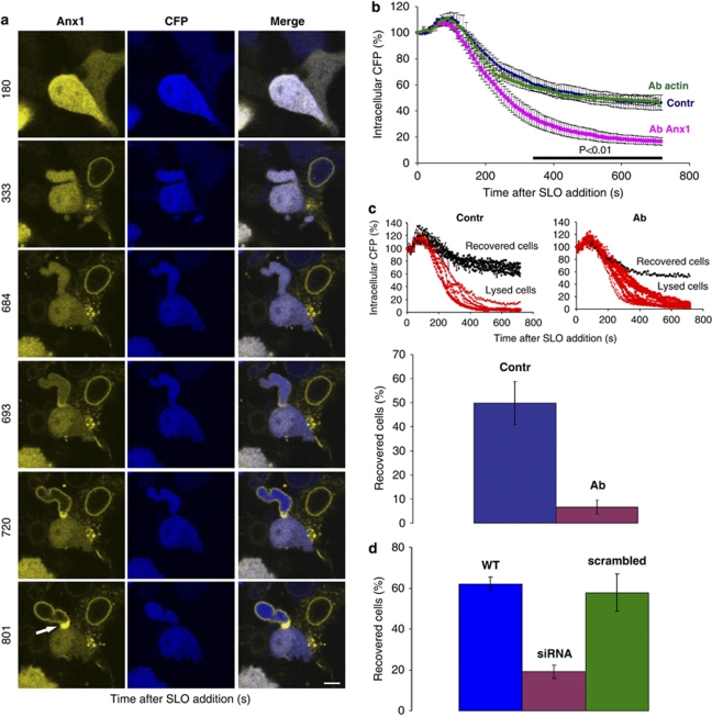
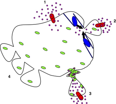
The plasma membrane constitutes a barrier that maintains the essential differences between the cytosol and the extracellular environment. Plasmalemmal injury is a common event during the life of many cells that often leads to their premature, necrotic death. Blebbing - a display of plasmalemmal protrusions - is a characteristic feature of injured cells. In this study, we disclose a previously unknown role for blebbing in furnishing resistance to plasmalemmal injury. Blebs serve as precursors for injury-induced intracellular compartments that trap damaged segments of the plasma membrane. Hence, loss of cytosol and the detrimental influx of extracellular constituents are confined to blebs that are sealed off from the cell body by plugs of annexin A1 - a Ca(2+)- and membrane-binding protein. Our findings shed light on a fundamental process that contributes to the survival of injured cells. By targeting annexin A1/blebbing, new therapeutic approaches could be developed to avert the necrotic loss of cells in a variety of human pathologies.


Figures

Figure 1
Figure 1
An injury-induced influx of Ca2+ triggers blebbing, which is mediated by the contraction of myosin II. (a) HEK 293 cells sprout numerous blebs after SLO-induced perforation. The images of the CFP-expressing cells are presented in the grey-scale mode. Magnification bar=10 μm. (b) Bar graph depicting the percentage of cells manifesting blebs before the addition of SLO, after the addition of SLO, and after pre-treatment with blebbistatin followed by the addition of SLO. (c) The graph shows the percentage of SLO-perforated cells manifesting blebs at the indicated [Ca2+]free. Mean values (±S.E.M.) are presented
Figure 2
Figure 2
Blebbing is a universal cellular response to plasmalemmal injury. Blebbing was recorded in SLO-perforated primary cultures of human myometrial smooth muscle cells, Jurkat T cells and Raji B cells, in mechanically injured human astrocytoma cells (U 373 cells), and in HEK 293 cells that had been treated with bacterial phospholipase C. Raji cells are shown at low magnification: blebbing cells are marked by asterisks. The images of non-transfected (Raji and Jurkat) or the CFP-expressing cells (smooth muscle, U 373, HEK 293) are presented in grey-scale mode. Magnification bar=10 μm
Figure 3
Figure 3
Blebbing protects cells from plasmalemmal damage. (a) Lysed cells are characterized by membrane-translocated annexin A1-YFP (Anx1). Cells recovering from an SLO attack (characterized by a cytoplasmic localization of annexin A1-YFP) protrude numerous blebs (arrows). Magnification bar=10 μm. (b) Augmented blebbing is associated with cells that are recovering from an SLO attack: 82±6% of the blebbing cells (n=85) recovered from an SLO attack, as opposed to 38±6% of the non-blebbing ones (n=140; t-test, P<0.01). Mean values (±S.E.M.) for nine independent experiments are presented. (c) Non-permeabilized HEK 293 cells (double-transfected with annexin A1-YFP and CFP) that had been treated with either acrylamide (AA; 241 cells; 9 independent experiments) or calyculin (203 cells; 9 experiments) were more prone to blebbing than untreated cells (control: 440 cells; 15 experiments; P<0.01 for both experimental conditions compared with the control). In non-permeabilized cells that had been treated with staurosporine (226 cells; 9 experiments), W7 (231 cells; 9 experiments) or blebbistatin (BS; 125 cells; 4 experiments), the inhibition was obscured by a low level of background blebbing. Mean values (±S.E.M.) are presented. (d) After pharmacological treatment the cells supplying the data in (c) were permeabilized with SLO. SLO-permeabilized cells that had been pre-treated with either acrylamide or calyculin were more prone to blebbing than untreated SLO-permeabilized cells. In contrast, pre-treatment with staurosporine, W7 or blebbistatin inhibited SLO-induced blebbing. Mean values (±S.E.M.) are presented (P<0.01 for any experimental condition compared to the control). (e, f) The cells supplying the data in (d) were analysed for their annexin A1 (e) or CFP (f) status. Mean values (±S.E.M.) are presented. In (e), the filled symbols denote the times after SLO exposure at which the differences between any experimental condition and the control reached statistical significance (t-test; P<0.01). In (f), the horizontal bar denotes the times after SLO exposure at which the differences between any experimental condition reached statistical significance (t-test; P<0.05)
Figure 4
Figure 4
Blebbing protects injured cells by isolating the damaged regions. Blebbing, an elevation in [Ca2+]i (membrane translocation of annexin A1) and the loss of cytoplasm (CFP-leakage) were simultaneously monitored in SLO-permeabilized HEK 293 cells (a, b) and in mechanically injured U 373 cells (c). Numbers and arrows highlight the regions of interest that are discussed in the text. Magnification bar=10 μm
Figure 5
Figure 5
Intrableb elevation in Ca2+. SLO was added (time=0) to non-transfected HEK 293 cells that had been pre-loaded with Fluo 5F. At time points=90, 108 and 117 s, [Ca2+] was higher in the bleb (arrow) than in the cytoplasm. Fluorescence is shown in the rainbow mode: low [Ca2+] – blue; high [Ca2+] – red. Magnification bar=10 μm
Figure 6
Figure 6
Annexin A1 blocks the communication between the damaged bleb and the rest of the cytoplasm. (a) A massive accumulation of annexin A1 is seen at the base of the SLO-perforated bleb (arrow). The cell in the upper right-hand corner is irreversibly perforated. Magnification bar=10 μm. (b) The SLO-induced loss of CFP recorded from a whole field of HEK 293 cells (containing minimally 15 cells) was significantly faster in the presence of the anti-annexin A1 antibody (Ab Anx1) than that either in the presence of the anti-β-actin antibody of the same isotype (Ab actin) or in the absence of antibody (Contr). Mean values (±S.E.M.) of three independent experiments. The horizontal bar indicates the times after SLO exposure at which the differences between experimental conditions (Ab Anx1 compared with either Ab actin or control) reached statistical significance (t-test; P<0.01). (c) Upper panels: assessed individually, SLO-permeabilized cells either completely lost their cytoplasm (lysed cells) or, after a partial loss of CFP, were able to repair the damaged plasma membrane and thus averted a further loss of cytoplasm (recovered cells). Representative experiments are shown. When perforated in the presence of the anti-annexin A1 antibody (Ab), only 6.6±2.9% of the cells (lower panel; 99 individual cells, n=5) recovered from the SLO attack, whereas 49.8±8.9% (lower panel; 73 individual cells, n=5) recovered from the perforation in the absence of the antibody (Contr; t-test P<0.01). Mean values (±S.E.M.) are represented. (d) SLO-induced lysis was significantly accelerated in HEK 293 cells stably expressing annexin A1-specific siRNA (siRNA) compared with either wild-type HEK 293 or HEK 293 cells transfected with scrambled siRNA (WT: 292 individual cells, n=9; siRNA: 348 individual cells, n=12; scrambled siRNA: 125 individual cells, n=5; t-test P<0.01). Mean values (±S.E.M.) are presented
Figure 7
Figure 7
Blebbing confers resistance against cell lysis. (1) Plasmalemmal injury: injury – red cylinder; Ca2+ influx – pink circles. (2) Injury-induced bleb formation: myosin-driven contraction – black arrows; cortical cytoskeleton – blue; cytoplasmic annexin A1 – green ovals. (3) Plugged bleb: membrane-bound annexin A1 complexed with Ca2+ (green ovals+pink circles). (4) Spontaneously protruding blebs

Similar articles

Cited by

References

    1. McNeil PL, Steinhardt RA. Plasma membrane disruption: repair, prevention, adaptation. Annu Rev Cell Dev Biol. 2003;19:697–731. - PubMed
    1. McNeil PL, Kirchhausen T. An emergency responce team for membrane repair. Nat Rev Mol Cell Biol. 2005;6:499–505. - PubMed
    1. Walport MJ. Complement. Second of two parts. N Engl J Med. 2001;344:1058–1066. - PubMed
    1. Parker MW, Feil SC. Pore-forming protein toxins: from structure to function. Prog Biophys Mol Biol. 2005;88:91–142. - PubMed
    1. Keefe D, Shi L, Feske S, Massol R, Navarro F, Kirchhausen T, et al. Perforin triggers a plasma membrane-repair response that facilitates CTL induction of apoptosis. Immunity. 2005;23:249–262. - PubMed

Publication types