. 2010 Jul 1;35(13):2121-3.
doi: 10.1364/OL.35.002121.

Time-resolved diffuse optical tomography with patterned-light illumination and detection

Affiliations

Time-resolved diffuse optical tomography with patterned-light illumination and detection

Jin Chen et al. Opt Lett. .

Abstract

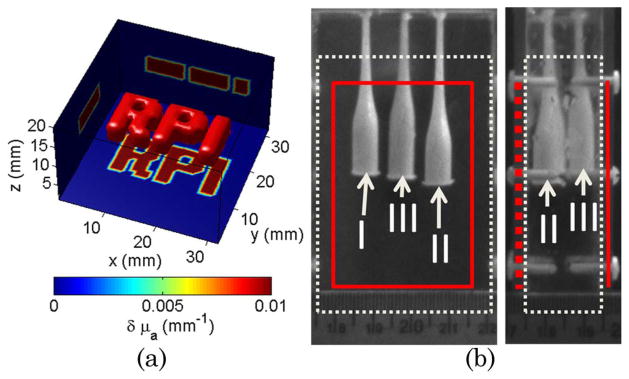
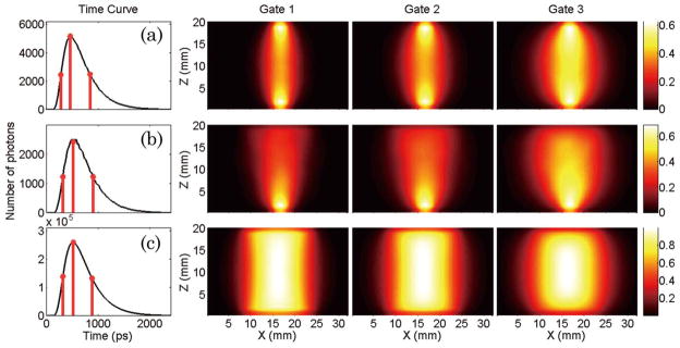
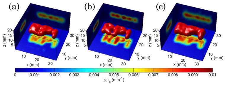
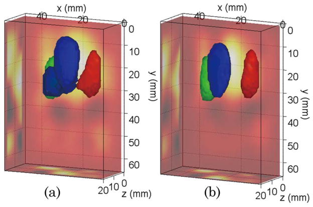
This investigation explores the feasibility of performing diffuse optical tomography based on time-domain wide-field illumination and detection strategies. Wide-field patterned excitation and detection schemes are investigated in transmittance geometry with time-gated detection channels. A Monte Carlo forward model is employed to compute the time-resolved Jacobians for rigorous light propagation modeling. We demonstrate both in silico and experimentally that reconstructions of absorption structures based on wide-field patterned-light strategies are feasible and outperform classical point excitation schemes for similar data set sizes. Moreover, we demonstrate that time-domain information is retained even though large spatial areas are illuminated. The enhanced time-domain data set allows for quantitative three-dimensional imaging in thick tissue based on relatively small data sets associated with much shorter acquisition times.


Figures

Fig. 1
Fig. 1
(Color online) (a) In silico phantom with complex absorptive structure. (b) Experimental phantom with three absorptive inclusions. The solid line (red) corresponds to the illuminated surface and point detector distribution. The dotted lines (white) delineate the reconstructed volume.
Fig. 2
Fig. 2
(Color online) Simulated detector readings for the central source–pattern and associated Jacobians for the three gates selected: (a) point source–point detector (• + •), (b) patterned illumination–point detector (□ + •), and c) patterned illumination–patterned detection (□ + ⊡).
Fig. 3
Fig. 3
(Color online) Optical reconstructions of the phantom of Fig. 1(a) for (a) point source–point detector time-gated data, (b) patterned excitation–point detector, and (c) patterned excitation–patterned detector. The isovolume was set at 40% of the maximum reconstructed value. The projections of the central reconstructed plane are provided on each boundary for each case.
Fig. 4
Fig. 4
(Color online) Optical reconstructions of the phantom of Fig. 1(b) for the pattern source–point detector strategy: (a) CW data and (b) TG data. The isovolume was set at 50% of the maximum reconstructed value. The ray–sum projections are provided on the sides of the reconstructed volume.

References

    1. Intes X, Djeziri S, Ichalalene Z, Mincu N, Wang Y, St-Jean P, Lesage F, Hall D, Boas D, Polyzos M, Fleiszer D, Mesurolle B. Acad Radiol. 2005;12:934.
    1. Tromberg BJ, Pogue BW, Paulsen KD, Yodh AG, Boas DA, Cerussi AE. Med Phys. 2008;35:2443. - PMC - PubMed
    1. Strangman G, Boas DA, Sutton JP. Biol Psychiatry. 2002;52:679. - PubMed
    1. Intes X, Chance B. Radiol Clin N Am. 2005;43:221. - PubMed
    1. Cuccia DJ, Bevilacqua F, Durkin AJ, Ayers FR, Tromberg BJ. J Biomed Opt. 2009;14:024012. - PMC - PubMed

Publication types