. 2010 Jan 1;4(1):20-31.
doi: 10.2174/187231310790226206.

Chemical Tumor Biology of Heparan Sulfate Proteoglycans

Affiliations

Chemical Tumor Biology of Heparan Sulfate Proteoglycans

Karthik Raman et al. Curr Chem Biol. .

Abstract

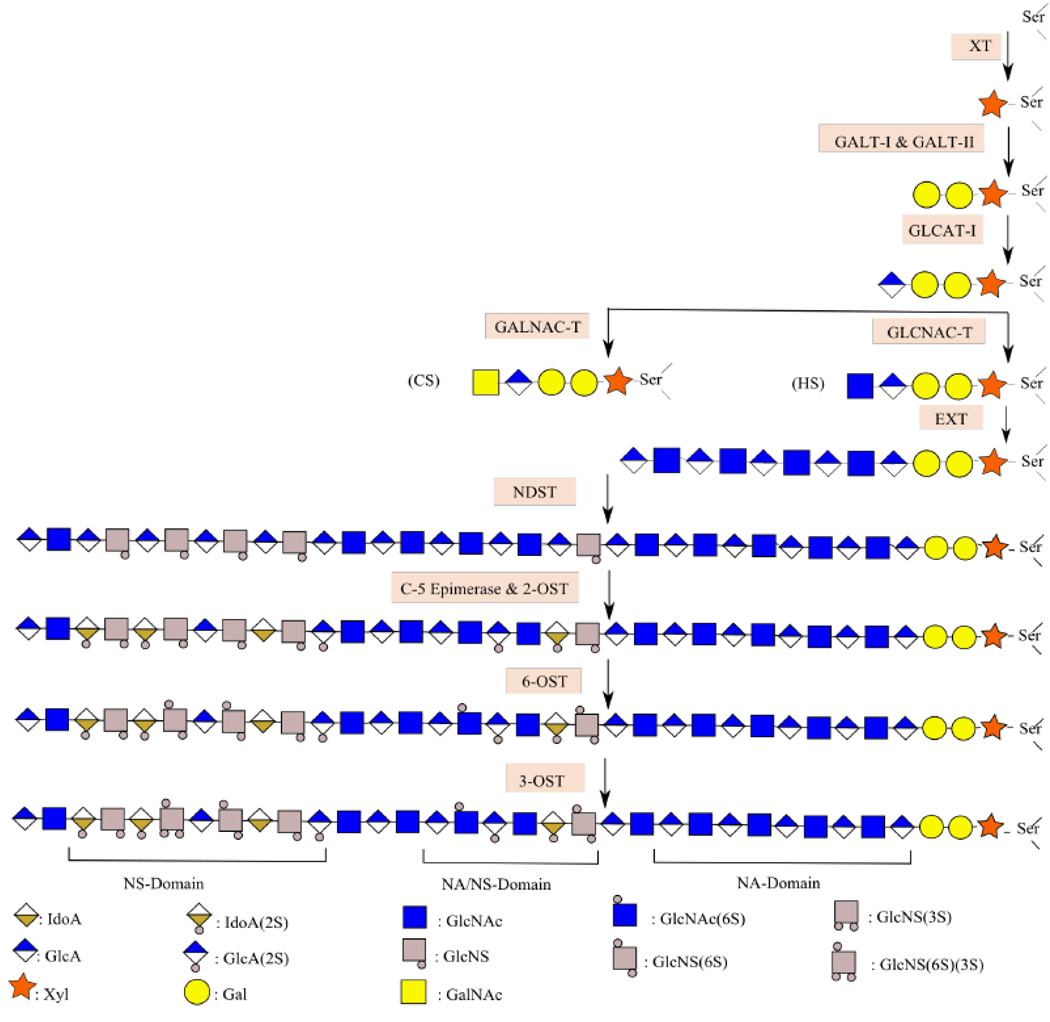
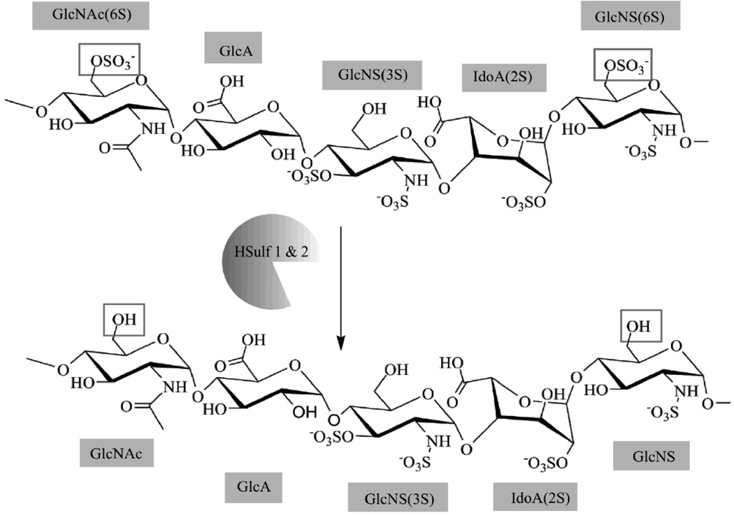
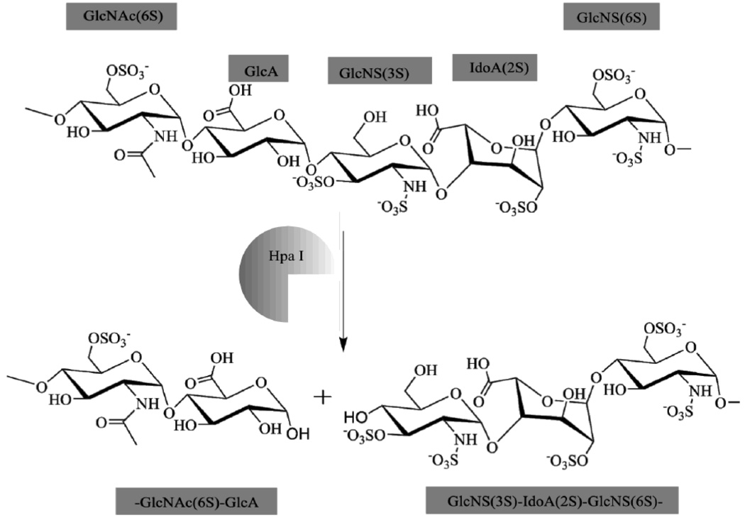
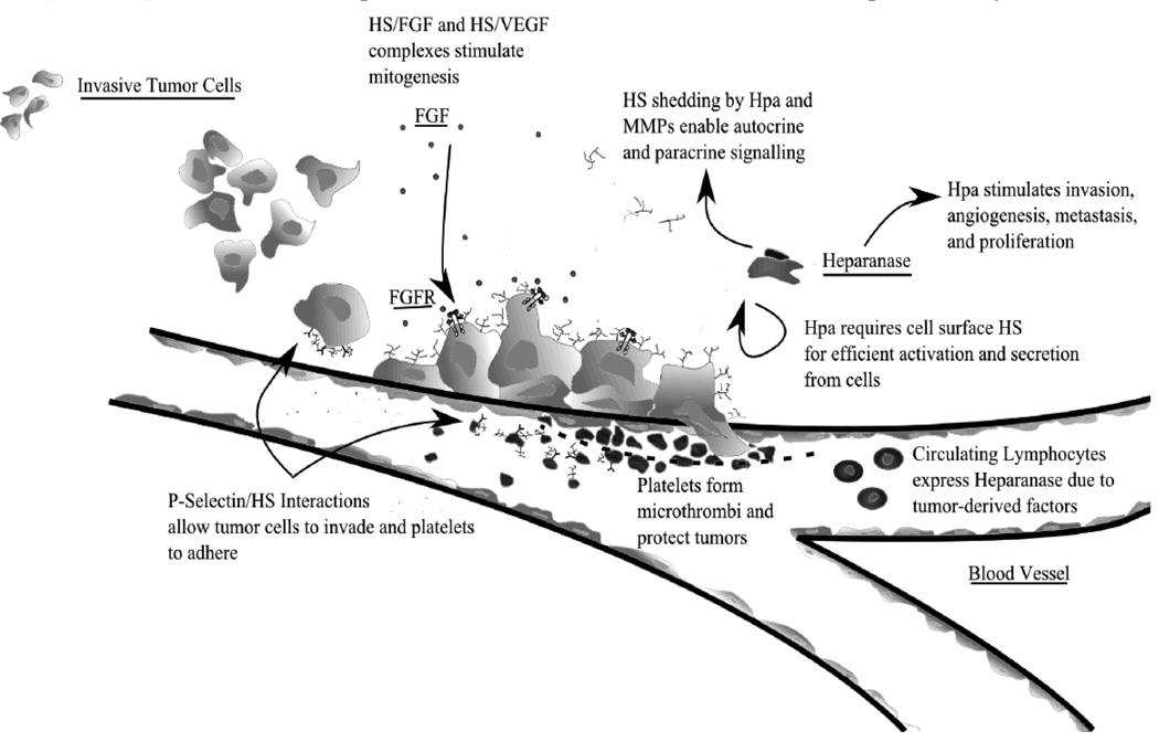
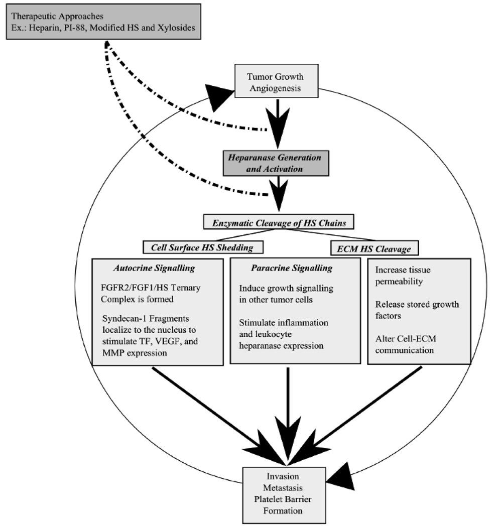
Heparan sulfate proteoglycans (HSPGs) play vital roles in every step of tumor progression allowing cancer cells to proliferate, escape from immune response, invade neighboring tissues, and metastasize to distal sites away from the primary site. Several cancers including breast, lung, brain, pancreatic, skin, and colorectal cancers show aberrant modulation of several key HS biosynthetic enzymes such as 3-O Sulfotransferase and 6-O Sulfotransferase, and also catabolic enzymes such as HSulf-1, HSulf-2 and heparanase. The resulting tumor specific HS fine structures assist cancer cells to breakdown ECM to spread, misregulate signaling pathways to facilitate their proliferation, promote angiogenesis to receive nutrients, and protect themselves against natural killer cells. This review focuses on the changes in the expression of HS biosynthetic and catabolic enzymes in several cancers, the resulting changes in HS fine structures, and the effects of these tumor specific HS signatures on promoting invasion, proliferation, and metastasis. It is possible to retard tumor progression by modulating the deregulated biosynthetic and catabolic pathways of HS chains through novel chemical biology approaches.


Figures

Fig. 1
Fig. 1. Proteoglycan Biosynthesis
Biosynthesis begins with the addition of xylose to serine residues of the core protein. Subsequently, glycosyl transferases add two Gal residues and one GlcA residue. EXT-1 and EXT-2 then extend the GAG chain by alternatively adding GlcNAc and GlcA. While elongation takes place, NDST-1 and NDST-2 convert certain GlcNAc to GlcNS residues. Epimerization and 2-O Sulfonation also occur in parallel; certain GlcA are converted to IdoA while 2-OST acts on certain GlcA and IdoA residues. Subsequent action of 6-OST and 3-OST yields the final PG chain that contains regions that have several N-Sulfated residues (NS Domain), others that have several N-Acetylated residues (NA Domain), and short regions that have a mixture of both (NA/NS Domain).
Fig. 2
Fig. 2. HSulf 1 & 2 Enzymatic Activity
HSulfs provide arylsulfatase activity selectively removing 6-O-Sulfate groups from GlcNAc and GlcNS residues. Deviant H-Sulf mRNA expression is present in several cancers.
Fig. 3
Fig. 3. Hpa I Enzymatic Activity
The Heparanase family is the only mammalian enzyme family that can cleave HS to form oligosaccharride units. Due to its unique activity, Hpa modulates growth factor signaling, ECM permeability and remodeling, cell clustering and adhesion, and several other cancer-related functions. Increased extracellular Hpa is an early indicator of malignant carcinomas.
Fig. 4
Fig. 4. The roles of HS in cancer
From gathered evidence presented in this article, HS is involved in several key functions regarding cancer progression. Cell metastasis depends on HS to provide P-Selectin binding so cells can adhere. As an antenna molecule, HS also plays a role in transducing signals from external growth factors by forming complexes with growth factor receptors. Additionally it is necessary for efficient activation of Heparanase, which is necessary for invasion, angiogenesis, and inflammation. By binding P-Selectins on platelets, HS also protects cancer cells from natural killer (NK) cell activity. Systemically, shed cell-surface HS can form paracrine signaling complexes FGFRs. Additionally, though not completely understood, cancer cells instigate circulating lymphocytes to upregulate heparanase activity; as an antenna molecule, HS is probably involved in this pathway as well.
Fig. 5
Fig. 5. Current HS-Based drug development strategies and their target signaling pathways in the Heparanase-induced self-proliferation loop
PI-88 and related oligosaccharides are potent anti-cancer agents because they interrupt Heparanase activity and also affect several growth signaling pathways such as the FGF2 pathway. Heparin and its low molecular weight anti-coagulant derivatives are natural Hep inhibitors that competitively inhibit Heparnase enzymatic activity by acting as the enzyme substrate. However, it is possible to affect both Heparanase as well as HS binding growth factors by utilizing modified HS and xylosides to tailor endogenous HS on cells to resist Hpa enzymatic activity and growth factor binding.
Fig. 6
Fig. 6. Chemical Structure of PI-88
A potent Heparanase inhibitor, PI-88 is a hyper-sulfated oligosaccharide that blocks Hpa and several HS/Growth factor interactions.

References

    1. Sasisekharan R, Shriver Z, Venkataraman G, Narayanasami U. Roles of heparan-sulphate glycosaminoglycans in cancer. Nat Rev Cancer. 2002;2:521–528. - PubMed
    1. Sanderson RD. Heparan sulfate proteoglycans in invasion and metastasis. Semin Cell Dev Biol. 2001;12:89–98. - PubMed
    1. Sanderson RD, Yang Y, Kelly T, MacLeod V, Dai Y, Theus A. Enzymatic remodeling of heparan sulfate proteoglycans within the tumor microenvironment: growth regulation and the prospect of new cancer therapies. J Cell Biochem. 2005;96:897–905. - PubMed
    1. Liotta LA, Kohn EC. The microenvironment of the tumour-host interface. Nature. 2001;411:375–379. - PubMed
    1. Carmeliet P. Angiogenesis in life, disease and medicine. Nature. 2005;438:932–936. - PubMed

LinkOut - more resources