Expression of stem cell factor/c-kit signaling pathway components in diabetic fibrovascular epiretinal membranes
- PMID: 20596251
- PMCID: PMC2893050
Expression of stem cell factor/c-kit signaling pathway components in diabetic fibrovascular epiretinal membranes
Abstract
Purpose: Stem cell factor (SCF)/c-kit signaling promotes recruitment of endothelial progenitor cells and contributes to ischemia-induced new vessel formation. We investigated the expression of the components of this pathway, including c-kit, SCF, granulocyte colony-stimulating factor (G-CSF), endothelial nitric oxide synthase (eNOS), and the chemokine receptor CXCR4, in proliferative diabetic retinopathy (PDR) epiretinal membranes.
Methods: Membranes from eight patients with active PDR and 12 patients with inactive PDR were studied by immunohistochemistry.
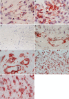
Results: Blood vessels expressed c-kit, SCF, G-CSF, eNOS, and CXCR4 in 18, 15, 19, 20, and 20 out of 20 membranes, respectively. Significant correlations were detected between the number of blood vessels expressing CD34 and the number of blood vessels expressing SCF (r=0.463; p=0.04), G-CSF (r=0.87; p<0.001), eNOS (r=0.864; p<0.001), and CXCR4 (r=0.864; p<0.001). Stromal cells expressed c-kit, SCF, eNOS, and CXCR4 in 19, 15, 20, and 20 membranes, respectively. The numbers of blood vessels expressing CD34 (p=0.005), c-kit (p=0.03), G-CSF (p=0.007), eNOS (p=0.001), and CXCR4 (p=0.018) and stromal cells expressing c-kit (p=0.013), SCF (p<0.001), eNOS (p=0.048), and CXCR4 (p=0.003) were significantly higher in active membranes than in inactive membranes.
Conclusions: SCF/c-kit signaling might contribute to neovascularization in PDR.
Figures


References
-
- Asahara T, Murohara T, Sullivan A, Silver M, van der Zee R, Li T, Witzenbichler B, Schatteman G, Isner JM. Isolation of putative progenitor endothelial cells for angiogenesis. Science. 1997;275:964–7. - PubMed
-
- Fazel SS, Chen L, Angoulvant D, Li S-H, Weisel RD, Keating A, Li R-K. Activation of c-kit is necessary for mobilization of reparative bone marrow progenitor cells in response to cardiac injury. FASEB J. 2008;22:930–40. - PubMed
-
- Huang PH, Chen YH, Wang CH, Chen JS, Tsai HY, Lin FY, Lo WY, Wu TC, Sata M, Chen JW, Lin SJ. Matrix metalloproteinase-9 is essential for ischemia-induced neovascularization by modulating bone marrow-derived endothelial progenitor cells. Arterioscler Thromb Vasc Biol. 2009;29:1179–84. - PubMed
-
- Li TS, Hamano K, Nishida M, Hayashi M, Ito H, Mikamo A, Matsuzaki M. CD117+ stem cells play a key role in therapeutic angiogenesis induced by bone marrow cell implantation. Am J Physiol Heart Circ Physiol. 2003;285:H931–7. - PubMed
-
- Miyamoto Y, Suyama T, Yashita T, Akimaru H, Kurata H. Bone marrow subpopulations contain distinct types of endothelial progenitor cells and angiogenic cytokine-producing cells. J Mol Cell Cardiol. 2007;43:627–35. - PubMed
Publication types
MeSH terms
Substances
LinkOut - more resources
Full Text Sources
Other Literature Sources
Medical
